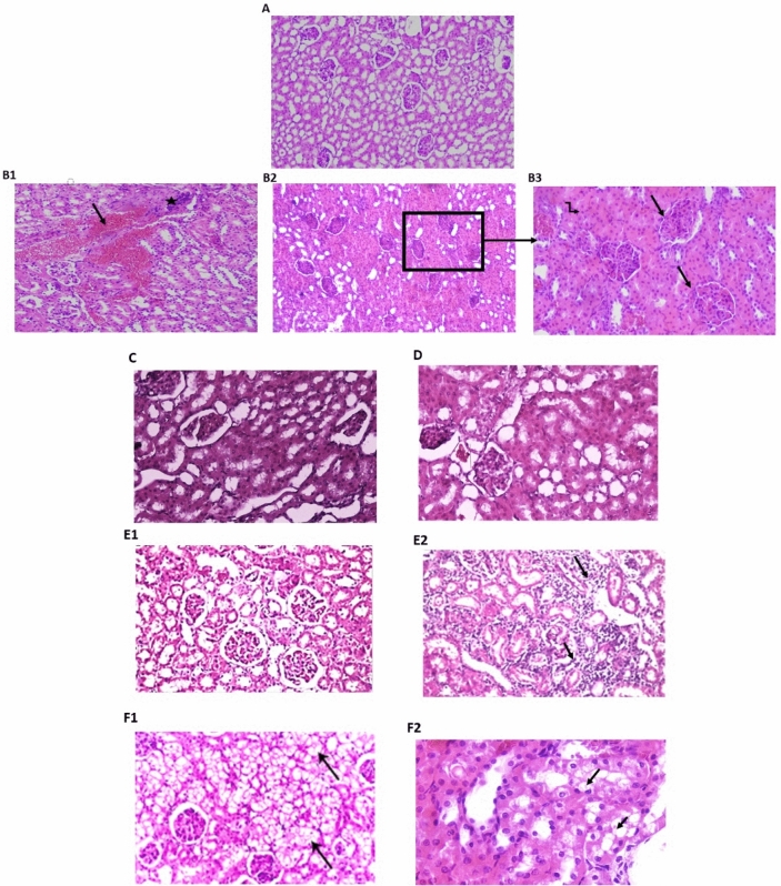
Figure 5.
Photomicrograph of kidney tissue sections in different studied groups. (H&E) (A) control group with normal histological structure of glomeruli and tubules (H&E ×200); (B1,B2) model group (dexamethasone-treated group) interstitial hemorrhage (arrow) ×100, hypercellular glomeruli (arrow) in (B3), ×400, obliterated tubules by swollen epithelial cells (zigzag arrow in B3); (C) PRV 0.5 mg/kg showing focally shrunken glomeruli and focally swollen hyalinized epithelial cells lining the tubules; (D) PRV1 mg/kg with well-formed glomeruli and tubules; (E) PRV 2 mg/kg exhibited a nearly normal histological renal pattern (E1, ×200) (arrows) with some inflammatory infiltrations (arrow E2); (F) losartan-treated group showing improved glomerular picture with interstitial vacuolation (F1, arrows), swollen hyalinized epithelial cells lining the tubules (arrows) were observed (F2, ×200).

