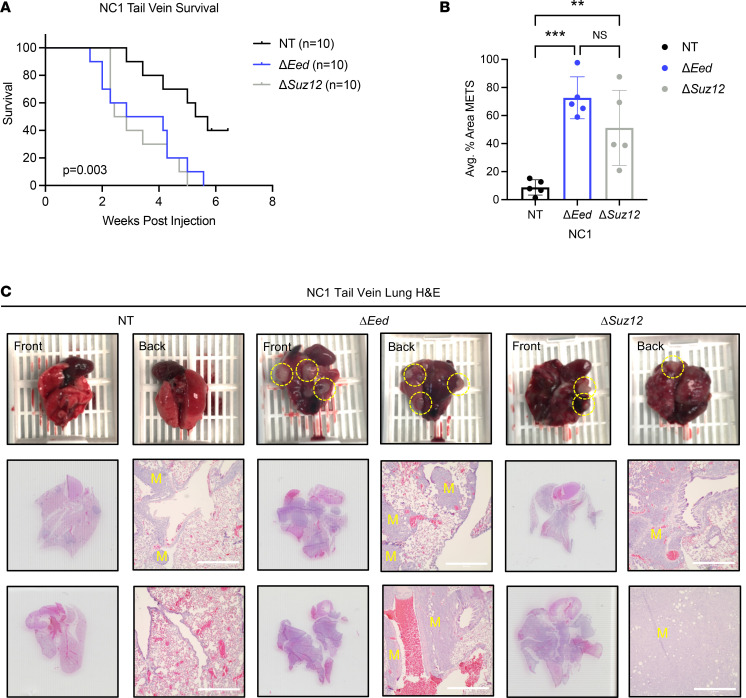
Figure 3. PRC2 loss increases metastatic colonization in vivo.
(A) Decreased survival of NSG mice receiving tail vein injections of NC1 cells with loss of Eed (n = 10) or Suz12 (n = 10) compared with NT (n = 10) cells with wild-type PRC2. (B) Increased metastatic lung area in mice receiving ΔEed (n = 5) and ΔSuz12 (n = 5) cells compared with NT (n = 5). Metastatic (MET) area quantification was performed on 5 lungs per group. Histological sections were taken at 0 μm, 100 μm, 200 μm, and 300 μm depths, and 4 images per section were taken at 4× objective. Data were analyzed using 1-way ANOVA with Tukey’s multiple comparisons. (C) Scans and 4× objective of 2 representative lungs from tail vein injections for each genotype. Metastases are indicated with a yellow “M.” Scale bars at 200 μm. Data represent biological replicates with the mean ± SD; **P < 0.01, ***P < 0.001.

