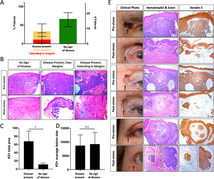
Fig 1. Keratin positive micro-tumors present despite complete clinical response to vismodegib.
A) Pathology results for excision samples after vismodegib treatment (error bars indicate 95% CI, n = 9 (left column), n = 18 (right column)), B) Pre- and post-vismodegib histology, C) Average total area and D) Average lesion size (pixels) of keratin 5 positive staining (normalized to section length, error bars indicate SEM, unpaired t-test *p<0.05, n = 7 (left column), n = 11 (right column)), E) Clinical photo, histology, and keratin staining pre and post-vismodegib treatment of patients scored as “no sign of disease” (black arrow–tumor location, yellow box—area of peripheral palisading).

