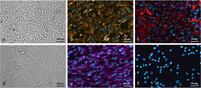
Fig 2. Characterization of human pleural mesothelial cells (HPMC) and human pleural fibroblasts (HPF).
Confluent cell cultures of human pleural mesothelial cells (HPMC) (a) and human pleural fibroblasts (HPF) (d). HPMC show polygonal appearance characteristic at confluency, whereas HPF are spindle-shaped cells, growing in parallel, whorl-forming arrays (phase-contrast microscopy; original magnification, x10). Immunofluorescent staining of fixed human mesothelial cells and fibroblasts with (b) monoclonal anti-vimentin and (c) monoclonal anti-cytokeratin for HPMC, x20 and (e) monoclonal anti-PHD1 and (f) monoclonal anti-cytokeratin for HPF, controls where primary antibody has been omitted do not show any unspecific binding, Scale bar = 50 and 100 μm.

