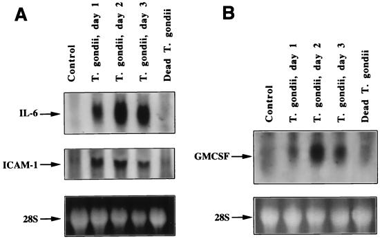
FIG. 2.
Northern blot analyses of the expression of IL-6 and ICAM-1 (A) and GM-CSF (B) mRNA in T. gondii-infected HRPE cells. Total cellular RNA isolated from HRPE cells at indicated time points were subjected to Northern blot analysis. The arrows indicate the positions of IL-6 (1.3 kb), ICAM-1 (3.3 kb), and GM-CSF (0.8 kb) mRNA. Lanes: control, uninfected cells incubated for 3 days; T. gondii, days 1, 2, and 3, cultures infected with T. gondii and total RNA prepared 1, 2, and 3 days p.i., respectively; dead T. gondii, cells incubated for 3 days with heat-killed T. gondii. The bottom panel shows the intensity of the 28S RNA band of an ethidium bromide-stained RNA gel.

