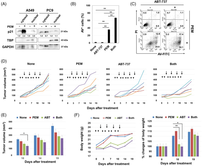
FIGURE 2.

Anti‐tumour effects of PEM and/or ABT‐263 in A549 and A549‐KOp21 cells in vitro and in vivo. (A) Cells were treated with PEM (1 μM) for 48 h. After harvesting, the cytoplasmic and nuclear fractions were separated, and immunoblotting was performed. The band density of cytoplasmic and nuclear p21 was normalized to GAPDH and TBP, respectively. (B) A549 cells were treated with PEM (1 μM) for 48 h. After harvesting, the collected cells were cultured with ABT‐737 (2.5 μM) for an additional 48 h. Thereafter, an apoptosis assay was performed. Data are the means ± SD of three replicates. **p < 0.01. (C) Representative results are shown. The numbers are the percentages of each subset. (D) BALB nude mice were injected with A549 (2 × 10⁶) cells and Matrigel into the right flank. When the tumour diameter was approximately 5–6 mm, PEM (100 mg/kg) (arrowheads) was i.p. injected on Days 0, 1, 3, 4, 6, 7, 9 and 10 after grouping. ABT‐737 (50 mg/kg) (arrows) was i.p. injected on Days 2, 5, 8 and 11 after grouping. Lines represent the growth of individual mice. Each group included 5–6 mice. (E) Tumour sizes are shown. *p < 0.05. (F) Body weights are shown. *p < 0.05, **p < 0.01. PEM, pemetrexed.
