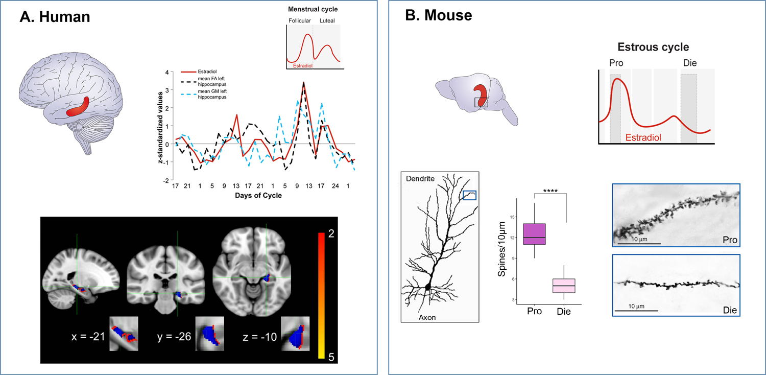
Figure 4. Comparable changes in hippocampal brain structure across the menstrual cycle in humans and the estrous cycle in mice.

A. In the human hippocampus (red), bilateral fractional anisotropy and left grey matter values are plotted against estrogen levels (in red) across the two menstrual cycles assessed (upper panel; adapted from Barth et al, 2016). In the lower panel, grey matter changes in left hippocampus are displayed with blue voxels corresponding to statistically significant results. B. In the mouse hippocampus (red), we showed structural changes in the ventral hippocampus (black square) as estradiol levels vary across the estrous cycle. In the lower panel, dendritic spine density is shown in the high-estrogenic (Pro, proestrus) phase versus low-estrogenic (Die, diestrus) phase (adapted from Jaric et al, 2019b).
