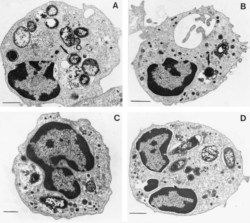
FIG. 2.
Increased number of E. coli seen in the cytoplasm of transmigrated PMNs under electron microscopy. (A) Transmigrated PMNs. Numerous bacteria are noted in the cytoplasm of PMNs (arrow). (B) Control PMNs. A few bacteria are observed in the cytoplasm of PMNs incubated for 120 min with bacteria in HBSS with calcium and 10−7 M fMLP (arrow). (C) Transmigrated PMNs in the presence of anti-CD11b antibody. (D) Transmigrated PMNs in the presence of anti-CD47 antibody. Bars, 10 μm.

