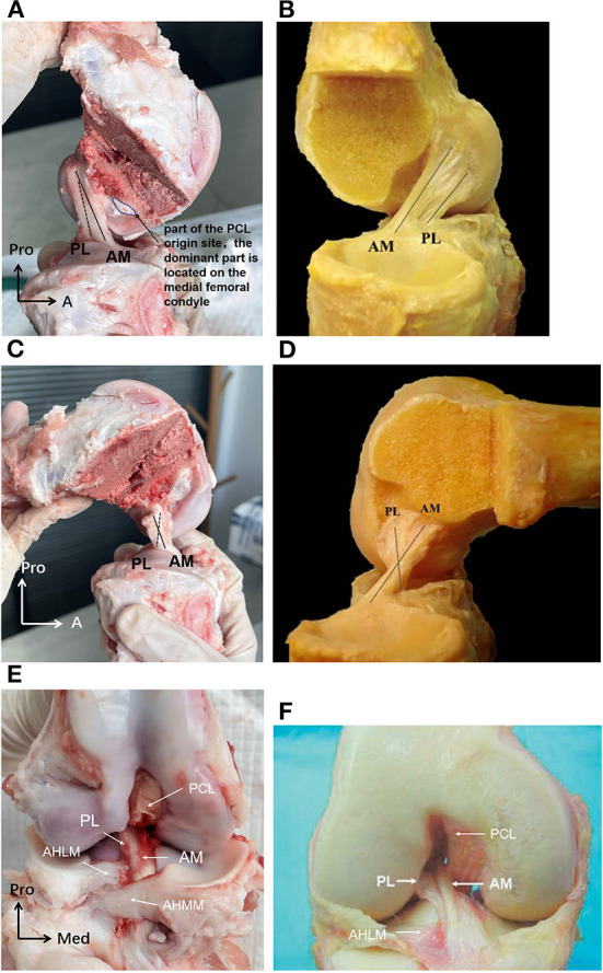
Figure 5.

(A) The porcine ACL at full knee extension in sagittal view. The AM bundle was lax while the PL bundle was tight, and the two bundles were almost parallel. (B) The human ACL at full knee extension in sagittal view. (C) The porcine ACL with knee flexed to around 90. The AM bundle was tight while the PL bundle was lax, and the two bundles formed an intersecting structure. (D) The human ACL with knee flexed to around 90. (E) The porcine ACL in the coronal plane. The AM bundle of the porcine ACL was anterior and medial to the PL bundle at the tibial insertion. The AHLM (anterior horn of the lateral meniscus) split the porcine ACL into AM and PL bundles. (F) The human ACL in the coronal plane. The AM bundle of the human ACL was anterior and medial to the PL bundle at the tibial insertion. The AHLM borders on the lateral side of the human ACL (23).
