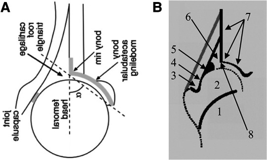
Figure 2.

Schematic of the systematic evaluation of hip sonograms of different original articles (A). The original diagram from (5) was rotated by 90° and mirrored around the transversal axis for better clarity (ultrasound from the left, upright image). (B) Scheme of anatomical identification (Checklist I) from a course manual for the ultrasound course according to Graf (6). The numbers describe: 1, ChB (chrondro-osseous border); 2, femoral head; 3, synovial fold; 4, joint capsule; 5, labrum; 6, cartilage; 7, bony roof; 8, bony rim (turning point).
