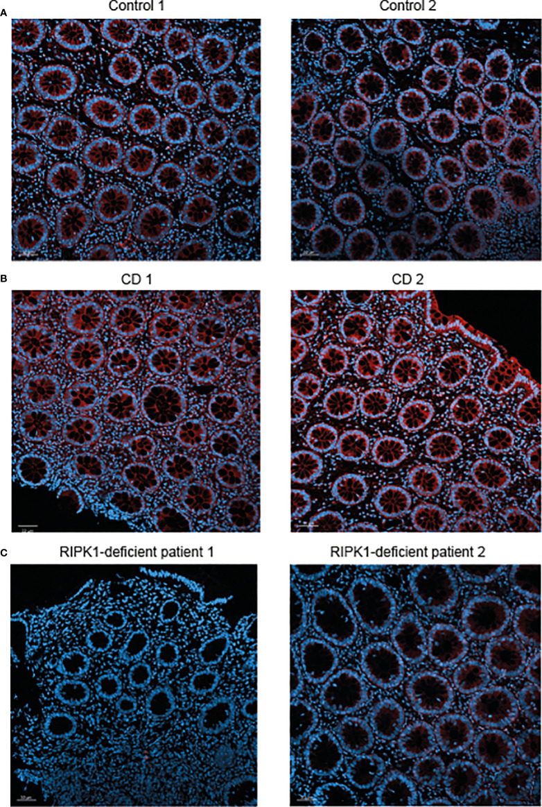
Figure 2.
Reduced RIPK1 expression in rectal samples of RIPK1-deficient patients. Figure depicts RIPK1 Immunofluorescence microscopy staining of rectal FFPE sections from (A) two control subjects, (B) two patients with active Crohn’s disease and (C) the two studied patients with RIPK1 deficiency. Sections were stained with anti-RIPK1 antibody (red) and counterstained with DAPI (blue). Magnification 20× (scale bar = 50 µm).

