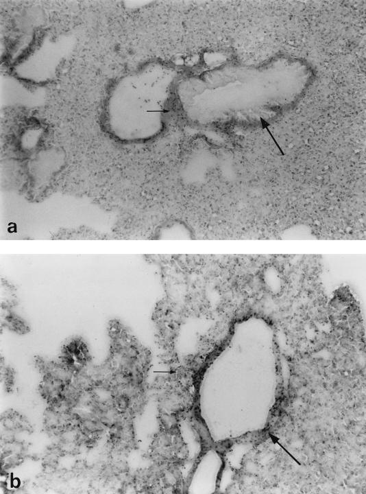
FIG. 4.
Light microscopy of lung tissue from mice infected with 106 CFU of S. pneumoniae PLN-A, sacrificed at 24 h (a) (the large arrow indicates a bronchiole, and the small arrow indicates slight cellular infiltration) and 48 h (b) (the large arrow indicates a bronchiole, and the small arrow indicates cellular infiltration) postinfection. Magnifications, ×300 (a) and ×400 (b).

