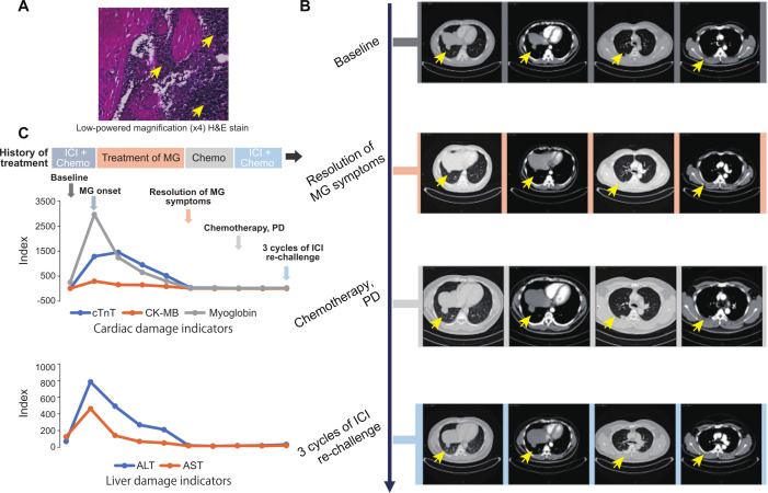
Figure 1.
Response to (re)immunotherapy and management of immune-related MG. (A) Low-powered magnification (×4) H&E stain of the thymic lesion. The yellow arrows indicate the thymic lesion. (B) Representative CT images of patients during the treatment (from the baseline to ICI rechallenge). The yellow arrows indicate the thymic lesion. (C), Change in levels of blood indicators of cardiac and liver damage. The arrows with different color represent the indicated stage of the treatment. ALT, alanine aminotransferase; AST, aspartate transaminase; cTnT, cardiac troponin T; CK-MB, creatine kinase-MB; ICI, immune checkpoint inhibitor; MG, myasthenia gravis.

