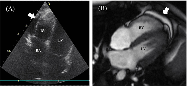
Figure 1. Echocardiogram and cardiac four-chamber cine magnetic resonance image.
(A) Echocardiogram obtained on the 33rd day after a second COVID-19 messenger RNA vaccination. Pericardial effusion is observed at the right ventricular side. (B) Cardiac four-chamber cine magnetic resonance image obtained on the 46th day after vaccination. Pericardial effusion is observed, but no abnormalities in the myocardium were found
RA: right atrium; RV: right ventricle; LV: left ventricle

