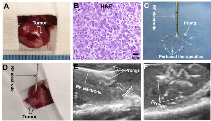
Figure 2.
Creation of rat orthotopic hepatic cancer for radiofrequency hyperthermia-enhanced treatment. (A) At 7 days after tumor implantation, an orthotopic hepatic tumor (arrow) is created in the rat liver. (B) Tumors were harvested 2 weeks after the treatments. H&E staining confirms the hepatocellular carcinomas formation (obtained from the controlled group). (C) Tumors received the treatments at 7 days after tumor implantation. The multimodal perfusion-thermal radiofrequency (RF) ablation electrode permits simultaneous peritumoral infusion of therapeutics and generation of RF hyperthermia. (D–F) Under real-time ultrasound imaging guidance, the multimodal ablation electrode is placed into the tumor and then multiple prongs are precisely deployed to cover the entire tumor to deliver therapeutics, followed by intratumoral radiofrequency hyperthermia to further enhance the treatment efficacy.

