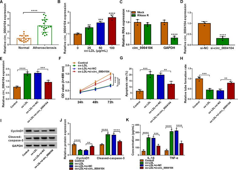
Fig. 2.
Circ_0004104 knockdown boosted proliferation and inhibited inflammatory in ox-LDL-exposed HUVECs. A Circ_0004104 level was determined in blood samples from 21 atherosclerosis patients and 17 healthy volunteers using qRT-PCR. B Circ_0004104 expression was measured in HUVECs treated with 0 μg/mL, 25 μg/mL, 50 μg/mL and 100 μg/mL of ox-LDL using qRT-PCR. C qRT-PCR analysis of circ_0004104 expression in HUVECs treated with or without RNase R assay. D qRT-PCR analysis of circ_0004104 expression HUVECs transfected with si-NC or si-circ_0004104. (E–J) HUVECs were treated with Control, ox-LDL, ox-LDL + si-NC, or ox-LDL + si-circ_0004104. E qRT-PCR analysis of circ_0004104 expression in treated HUVECs. F Cell proliferation was assessed in treated HUVECs using MTT assay. G Apoptosis rate was analyzed in treated HUVECs using flow cytometry assay. H The ability of HUVEC tube formation was assessed using tube formation assay. I and J Protein levels of CyclinD1 and Cleaved-caspase-3 in treated HUVECs were detected using western blot assay. K The secretion of inflammatory factors IL-1β and TNF-α in treated HUVECs was analyzed using ELISAs. **P < 0.01; ***P < 0.001; ****P < 0.0001. All cellular experiments were independently repeated three times. Student’s t-test was used to analyze the differences in (A and D), one-way ANOVA was utilized to assess the differences in (F, I, and K), and two-way ANOVA was utilized to analyze the differences in (B, C, G, and H)

