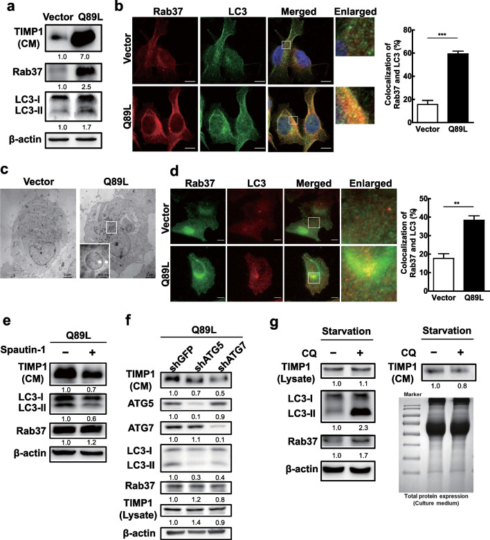
Fig. 3.
Active-form Rab37-mediated TIMP1 secretion was associated with increased autophagic activity and colocalization of Rab37 and LC3. a Lung cancer CL1-5 cells stably expressing active-form Rab37 (Q89L) or (Vector) plasmid were cultured for 24 h. The levels of TIMP1 in the conditional medium and the proteins of active-form Rab37, total Rab37, LC3, and β-actin in the cell lysate were evaluated by immunoblotting using specific antibodies. β-Actin was used as the internal control. b CL1-5 Vector and Rab37 Q89L cells were maintained in the serum-free medium for 24 h. Rab37 protein was labeled by primary anti-hRab37 antibody and second antibody conjugated with rhodamine (red). LC3 protein was labeled by primary anti-LC3 antibody and second antibody conjugated with FITC (green). The labeled cells were observed under confocal microscopy. The quantification of colocalization of Rab37 and LC3 was conducted by randomly investigating 30 cells. c TEM was used to investigate the double membrane autophagosome-like vesicles in Vector and O89L cells. The white arrow points to the double membrane autophagosome-like vesicle. d CL1-5-Vector and CL1-5-Q89L cells were seeded on the slides for 24 h. The same procedure as in b was used to label Rab37 protein (green) and LC3 protein (red) and investigated under a total internal reflection fluorescence (TIRF) microscope. Scale bar = 10 μm. The quantification of colocalization of Rab37 and LC3 was conducted by randomly investigating 30 cells. e CL1-5-Q89L cells were treated with autophagy inhibitor spautin-1 (10 μM) for 24 h. The protein level of TIMP1 in the conditional medium, and LC3 and Rab37 proteins in the total cell lysate were evaluated by immunoblotting. f ATG5 and ATG7 genes were silenced by specific lentivirus shRNAs in CL1-5-Q89L cells. Lentiviral shGFP was used as the negative control. The protein levels of Atg5, ATG7, LC3, Rab 37, and TIMP1 in cell lysate, as well as TIMP1 in the conditional medium were evaluated by immunoblotting. g CL1-5-Q89L cells were treated without serum in the presence or absence of CQ (50 μM) for 24 h. The levels of TIMP1 in the conditional medium, total LC3, TIMP1, Rab 37, and β-actin in the cell lysate were evaluated by immunoblotting using specific antibodies. β-Actin was used as the internal control

