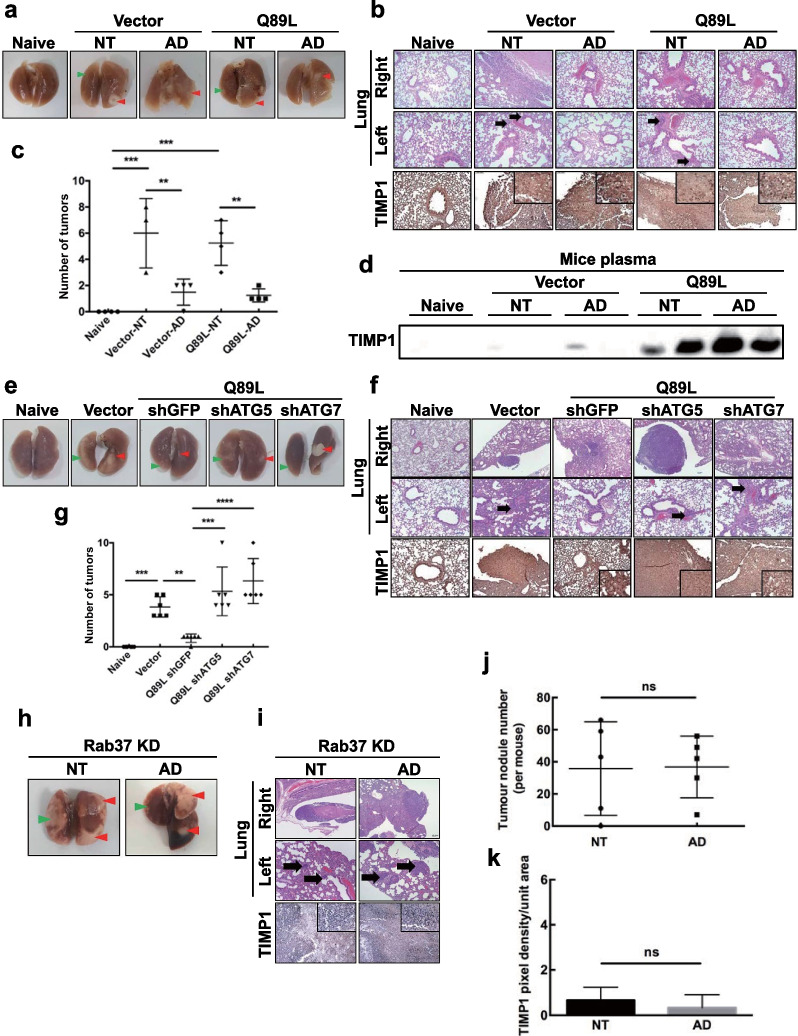
Fig. 5.
Autophagic promotes Rab37-mediated TIMP1 secretion and suppresses lung cancer cell metastasis in the lung-to-lung mouse metastasis model. Lung cancer CL1-5 control (Vector) and CL1-5-Q89L (Q89K) cells (5 × 10⁴ cells) were suspended in 10 μl of Matrigel. The Vector-AD and Q89L-AD group NOD-SCID mice received amiodarone (30 mg/kg) by i.p. injection twice a week for three weeks, together with the Naïve control group were sacrificed on day 35. Solid nodules located in the right lateral lung and within the pleural thoraxes were removed. a Five representative images of lungs were from the mice receiving CL1-5 Vector or CL1-5-Q89L cells with or without amiodarone together with the Naïve mice. Red arrow: primary tumor in the right lung; Green arrow: metastatic nodule in the left lung. b Row 1 and 2: H&E staining of lung tissues at local and distant regions of five groups. Row 3: IHC staining of the level of TIMP1 in the right lung tissues using anti-TIMP1 antibody. Scale bar = 10 μm. c Quantification of the lung tumors in the five groups of mice. d The five groups of NOD-SCID mice were inoculated i.p. with a single dose of amiodarone (30 mg/kg). Plasma samples were collected on the day of sacrifice, and TIMP1 protein level was measured by immunoblotting analysis using anti-TIMP1 antibody. Lung cancer Vector, CL1-5-Q89L-shGFP, -shAtg5, -shAtg7, and Naïve cells (5 × 10⁴ cells) suspended in 10 μl of Matrigel were inoculated into the five groups of mice (five mice/group). The solid nodules located in the right lung and within the pleural thoraxes were removed. e Five representative images of lungs were from the mice of the Naïve group and the mice receiving CL1-5-Q89L cells expressing Vector, -shGFP, -shAtg5, and -shAtg7 genes. Red arrow: primary tumor in the right lung; Green arrow: metastatic nodule in the left lung. f H&E stain showing tumor formation at the primary site (Row 1) and the distant regions (Row 2) in groups of Vector, Q89L-shAtg5, and Q89L-shAtg7 compared to the Q89L-shGFP group. Row 3: IHC stain showing the level of TIMP1 in the right lung tissues using anti-TIMP1 antibody. g Quantification of tumors in each group of mice. The NOD-SCID mice were divided into two groups (five mice/group). Lung cancer H460-shRab37 stable cells (5 × 10⁴ cells) suspended in 10 μl of Matrigel were inoculated into the right lung of the mice. The Rab37KD-AD group received amiodarone (30 mg/kg) by i.p. injection twice a week for two weeks. Mice were then sacrificed on day 35. The nodules located in the injected right lung and within the pleural thoraxes were removed. h Two representative images of lung tissues were inoculated with H460-shRab37 cells with (AD) or without amiodarone (NT). Red arrow: primary tumor in the right lung; Green arrow: metastatic nodule in the left lung. i Row 1 and 2: H&E staining of lung tissues at local and distant regions of the mice inoculated with H460 Rab37KD cells with (AD) or without amiodarone (NT). Row 3: IHC stain showing the level of TIMP1 in the left lung tissues using anti-TIMP1 antibody. Scale bars = 10 μm. j Quantification of tumors in the two mice groups. k Quantification of TIMP1 level in the two mice groups

