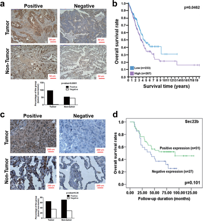
Fig. 7.
Analyses of significance of p62 accumulation and Sec22b level in clinical lung cancer specimens and an online database. a The sections of 103 lung cancer patient specimens were labeled by anti-p62 antibody followed by DAB staining (brown). Hematoxylin (blue) was used to stain the nuclei. Quantification of p62 level in lung cancer specimens is shown as a bar graph. Data were analyzed by chi-square test. Scale bar = 50 µm. b The survival rate of lung adenocarcinoma (LUAD) patients in an online database (n = 500) was calculated using Kaplan–Meier analysis and log-rank test (THE HUMAN PROTEIN ATLAS, https://www.proteinatlas.org/). c The sections of 136 lung cancer patient specimens were labeled by anti-Sec22b antibody. Quantification of Sec22b expression in lung cancer specimens is shown as a bar graph. Data were analyzed by chi-square test. Scale bar = 50 µm. d The survival rate of the lung cancer patients was determined by Kaplan–Meier survival analysis and log-rank test

