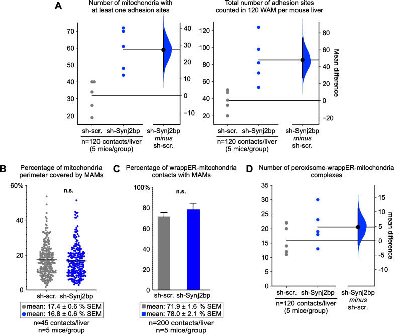
Fig. 5.
Synj2bp silencing in the mouse liver increases wrappER-mitochondria adhesion sites without affecting MAM content. A Quantitative EM morphometric analysis of wrappER-mitochondria adhesion sites. Here we counted the adhesion sites between the wrappER and the mitochondrial outer membrane in high-magnification EM images of wrappER-associated mitochondria (WAM) and processed the data by estimation statistics analysis [24, 25]. The graph shows that Synj2bp silencing increases the number of mitochondria with at least one adhesion site (left) and the number of adhesion sites counted in 120 WAM per liver (right; total n = 600 WAM; n = 5 mouse per condition). B, C Quantitative EM morphometric analyses of the MAMs present in wrappER-associated mitochondria. In B, each dot represents the length of a MAM expressed as a percentage of the perimeter length of the mitochondrion to which it belongs (measured in nm). This study shows that Synj2bp silencing changes neither the size of each MAM measured (B) nor the number of MAMs present in all WAMs analyzed (C). The Student’s t test was used to calculate p values. D Quantitative EM morphometric analysis of peroxisome-wrappER-mitochondria (PEWM) complexes in control and Synj2bp-silenced livers. Here we counted the PEWM complexes in high-quality EM liver images and processed the data by estimation statistics analysis. The graph shows that the number of PEWM complexes does not significantly change. Data were obtained using mouse livers at 3 h postprandial

