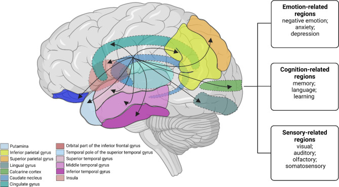
Fig. 5.
Schematic diagram of the CM-related functional brain connectivity network. The diagram shows our results about the significantly enhanced FC between the CN and brain regions associated with emotion, cognition, and sensory (the calcarine cortex, lingual gyrus, anterior cingulate cortex, superior and inferior parietal cortex, precuneus, temporal gyrus, insula, orbital part of the inferior frontal gyrus, and temporal pole of the superior temporal gyrus). The black arrows represent enhanced FC. The dotted line represents the tissue located inside the brain. Note: CM, chronic migraine; CN, caudate nucleus.

