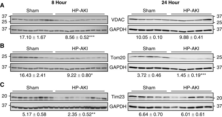
Figure 5.
Renal mitochondrial membrane integrity in sham and HP-AKI mice. Altered mitochondrial membrane integrity was observed at 8 and 24 hours after HP-AKI. (A) Decreased voltage-dependent anion channel (VDAC) protein expression was observed at 8 hours after HP-AKI, and (B) Tom20 protein expression was decreased at both 8 and 24 hours after HP-AKI. (C) Tim23 protein expression was significantly decreased at 8 hours after HP-AKI. Densitometry (mean±SEM), normalized for GAPDH expression, is displayed below blots. n=8 and n=10 in sham and HP-AKI groups, respectively, at 8 hours, and n=5 in each group at 24 hours. ***P<0.001, **P<0.01, and *P<0.05 versus sham group. At each time point (8 and 24 hours), analyses of VDAC and Tom20 were performed on the same membrane that was divided, as described in the Methods section. At each time point, the same GAPDH blot was employed for densitometric analysis for each of these targets and is displayed below both blots.

