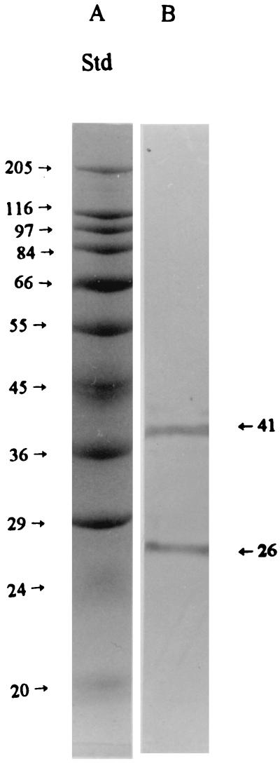
FIG. 5.
Typical immunoblotting of fimbrial binding proteins from the intestinal mucus of pigs. The mucus proteins were bound to the affinity column, and the column was successively washed with buffers A and B. The bound receptors were then eluted from the column with elution buffer (buffer C), and the purified receptor proteins (10 μl; 300 μg/ml) were applied to the electrophoresis gel in binding buffer (lane B). The proteins were separated by SDS-PAGE and electroblotted onto nitrocellulose membranes, and the receptors for fimbriae were stained by procedures described in Materials and Methods. Molecular mass standards (Std) are shown in lane A, with the corresponding molecular masses (kilodaltons) indicated to the left. The immunostaining pattern of the fimbrial binding receptors was the same when the receptors were bound to the column in binding buffer and directly eluted with buffer C (data not shown).

