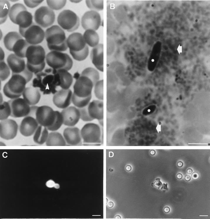
FIG. 3.
In vivo studies of the interactions between C. albicans ATCC 66369 (A) and C. krusei (B) and platelets. Blood smears were stained with MGG. Immunofluorescence (C) and phase-contrast (D) photographs of platelet adherence to C. parapsilosis blastoconidia are shown. Note blastoconidia of C. albicans (arrowhead) with a crown of platelets. Note blastoconidia of C. krusei (∗) associated with big aggregates of platelets (arrows). Bars, 10 μm.

