
Figure 6: Histopathology of proliferative diabetic vitreo-retinopathy (PDVR).
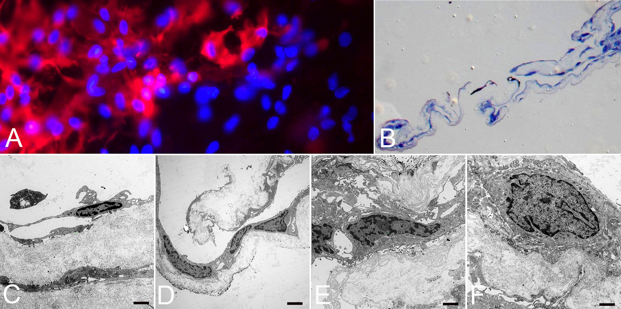
Immunofluorescence microscopy (A), light (B) and transmission electron microscopy (C-F) of a fibrocellular, premacular membrane surgically removed from an eye with PDVR. A) Flat-mounted membrane with anti-CD45 (red) positive staining merged with cell nuclei staining (blue) indicating the presence of hyalocytes (original magnification x400). B) Semithin section of membrane demonstrated folded fibrocellular composition with thick collagen strands (original magnification x100). C) Ultrastructural analysis revealed hyalocytes with elongated cell bodies and thin cell processes in abundance of native vitreous collagen (original magnification x7000, bar = 1000 nm). D) Hyalocytes and myofibroblasts situated on layers of vitreous collagen strands (original magnification x3000, bar = 2000 nm). E) Myofibroblasts surrounded by newly formed collagen (original magnification x7000, bar = 1000 nm). F) Fibroblast with large cell nucleus and newly formed collagen (original magnification x7000, bar = 1000 nm). Images courtesy of author R Schumann.