
Fig. 8.
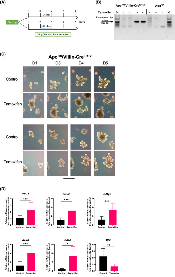
TRα1 modulation by the induction of the Apc mutation in mouse enteroids. (A) Schematic diagram of the protocol used for ex vivo enteroid cultures. (B) PCR analysis on gDNA extracted from enteroids of different genotypes and treatments, as indicated, to verify the recombination of the Apc gene after tamoxifen treatment. Specific primers recognizing the mutated allele were used. Note that the band corresponding to mutated Apc was detected only in Apc+/fl/villin‐CreERT2 tamoxifen‐treated organoids. (C) Bright‐field pictures of enteroids obtained from Apcf/+/villin‐CreERT2 and Apc+/fl mice treated with tamoxifen or not treated (control). Pictures were taken at different days of culture, as indicated, using a Zeiss AxioVert inverted microscope with a 10× objective. Scale bar = 10 μm. (D) RT‐qPCR analysis of the indicated genes performed on RNA isolated from Apc+/fl/villin‐CreERT2 enteroids treated with tamoxifen or not treated (control), as indicated. Histograms represent the mean ± SD (n = 6), and data are expressed as the fold change relative to the control condition (= 1). Ppib was used as a reference gene. *P < 0.05, **P < 0.01 and ***P < 0.001 by unpaired, two‐tailed Student's t‐test. The results in B–D are representative of three independent experiments, each conducted in six replicates.