
Figure 1. Oncolytic VVs elicit tumor-specific antitumor CD8+ T cell response in the tumor tissue.
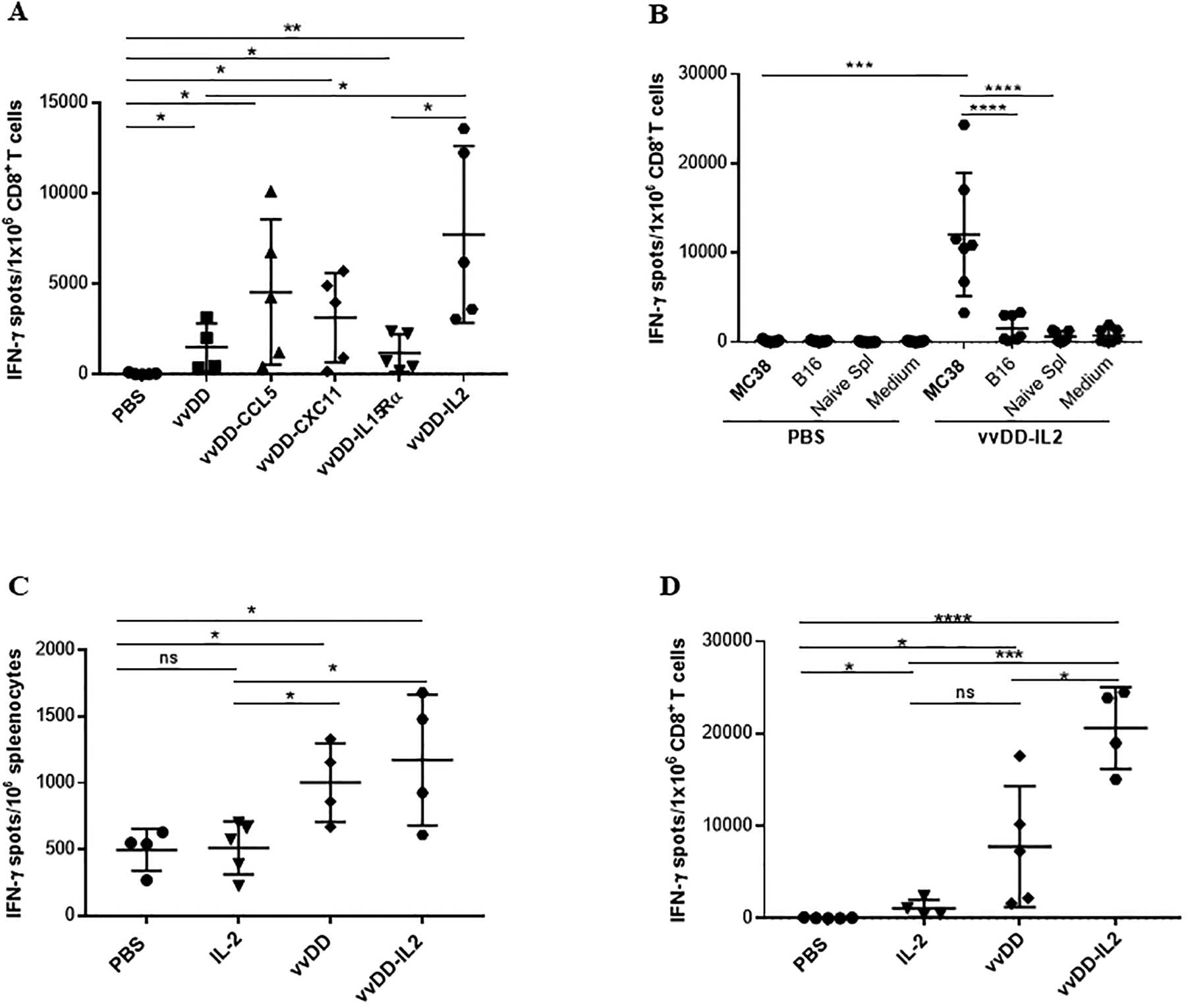
Ten days after viral treatments, subcutaneous MC38 tumors and/or splenocytes were collected and single cell suspensions were made followed by magnetic separation. Then isolated CD8+ T cells or splenocytes were tested by IFN-γ ELISPOT assay. (A). CD8+ T cells isolated from tumors (n = 4 – 5 mice/group) were either left unstimulated or challenged with γ-irradiated MC38 tumor cells for 24 h. Results were shown as individual data points (number of spots in each well) and bars (means ± standard deviation) of IFN-γ+ CD8+ T cells from each mouse evaluated in triplicate. Data from one experiment representing 2 independent experiments are shown. (B). Tumor-specificity of the OV-induced CD8+ TILs (n = 7 mice/group). Data are from one experiment representative of 3 independent experiments. For multiple group comparison, One-way ANOVA was used. ***p < 0.001; ****p < 0.0001. (C). MC38 tumor cell reactivity of splenocytes by IFN-γ ELISPOT assay. Splenocytes from treated mice were either left unstimulated or challenged with γ-irradiated MC38 tumor cells for 24 h. (D). MC38 tumor cell reactivity of isolated CD8+ TILs by IFN-γ ELISPOT assay. One experiment representative of 2 independent experiments is shown (n = 4–5 mice/group). Student’s t-test was used to analyze the statistical significance for data presented in panels A, C and D. *p < 0.05; **p < 0.01; ***p < 0.001; ****p < 0.0001.