
Figure 4. OV-induced TILs can be cultured and expanded ex vivo and retain their tumor specificity.
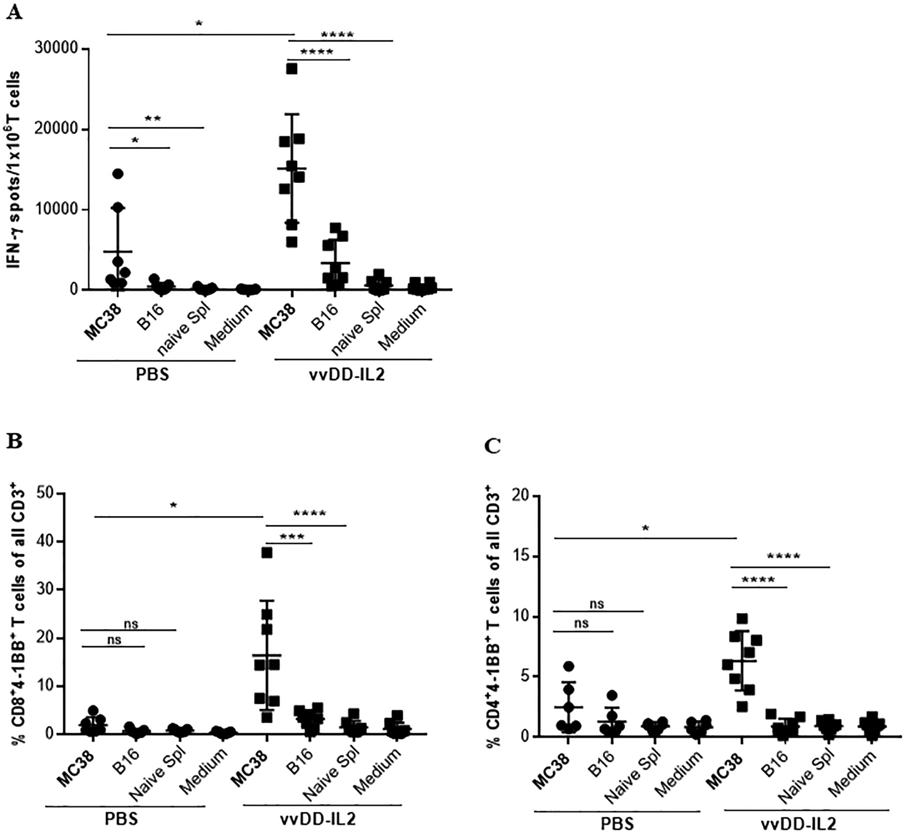
(A). Tumor specificity of the expanded TILs. TILs from each individual mouse have been cultured for 4 days and then tested by IFN-γ ELISPOT assay. (B, C). Analysis of 4–1BB upregulation on (B) CD4+ and (C) CD8+ T cells by flow cytometry. As previously described, T cells were either left unstimulated (medium) or challenged with γ-irradiated MC38 tumor cells or γ-irradiated B16 tumor cells or naïve splenocytes from non-tumor-bearing B6 mouse in duplicate. After 24 h the cells have been stained for flow cytometry analysis for CD3, CD4, CD8, 4–1BB. Results are shown as individual data points (percentage of CD8+4–1BB+ T cells and CD4+4–1BB+ T cells) and bars (means ± standard deviation) of T cells from each mouse evaluated in duplicate. Data are presented as summary from 2 out of 5 independent experiments (n = 3–4 mice/group). For multiple group comparison One-way ANOVA was used. *p < 0.05; **p < 0.01; ***p < 0.001; ****p < 0.0001.