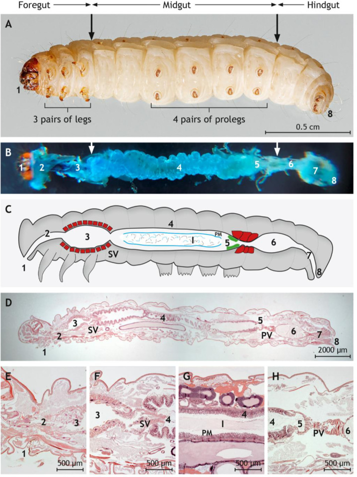
Fig 1. Anatomy and histology of G. mellonella larvae with emphasis on the digestive tract.
(A) Underside of a G. mellonella larva. The foregut, the midgut, and the hindgut are indicated by arrows. (B) Dissected digestive tract after instillation with methylene blue. (C) Schematic drawing of the digestive tract. The stomadeal valve (SV) separates foregut lined by cuticular epithelium from midgut lined by glandular epithelium. The proctodeal valve (PV) is located between midgut and hindgut. The distinct epithelium cranial to the PV is labeled in green. The ingesta (I) in the midgut is covered by the peritrophic membrane (PM) and separated from the mucosa by the ectoperitrophic space. The crop and both valves are surrounded by a thick layer of musculature (red). (D) Longitudinal and sagittal histological section along the middle through G. mellonella. (E) Magnification of mouth, esophagus, and crop lined by the cuticular epithelium and surrounded by muscle cells. (F) Magnification of the SV between crop and midgut. (G) Magnification of the midgut lined by glandular epithelium. The ingesta is surrounded by the peritrophic matrix (PM) and separated from mucosa by the ectoperitrophic space. (H) Magnification of the PV between midgut and hindgut lined by cuticular epithelium. Vacuolated columnar epithelial cells line the midgut cranial to the PV. (D)-(H) are paraffin sections stained by hematoxylin and eosin. Sections are indicated by numbers: 1 = mouth, 2 = esophagus, 3 = crop, 4 = glandular intestine, 5 = transition zone, 6 = cuticular intestine, 7 = rectum, 8 = anus. Photos of representative preparations are shown; the scales are indicated.

