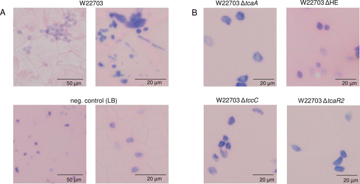
Fig 7. Hemocyte morphology 24 h p.i.
(A) Hemolymph cell preparations from G. mellonella orally infected with 6.1 × 10⁵ CFU of W22703 or treated with LB medium as control. (B) Hemocytes of larvae after application of four W22703 mutants, showing cell morphology similar to those of the controls. Hemocyte aggregation and deformation was visible only upon infection with W22703. Hemolymph preparations were fixed with methanol and stained by Giemsa solution. Photos of representative preparations are shown; cells vary in size, the scale is indicated. An Olympus BX53 microscope (Olympus Europa, Hamburg, Germany) was used.

