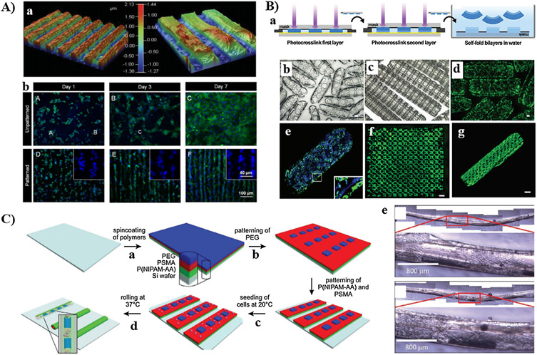
Fig. 12.
Integration of smart materials with photolithography: (A) Development of photopatterned GelMA hydrogel by spatial degradation of photocleavabie oNB ester linkages (a); DAPI (blue)/α-actinin (green) staining of cardiomyocytes cultured on non-patterned (top images) and patterned GelMA (bottom images) at days 1, 3, and 7 (b). (B) 4D biofabrication of self-folding PEG bilayers: (a) irradiation through photomask to crosslink the first and second PEG diacrylate layers of different molecular weights; self-folding of PEG bilayers into small (b) and large (c) cylindrical hydrogels with patterned holes; (d) calcein AM staining of fibroblast cells encapsulated within cylindrical cell-laden hydrogel; (e) Multi-culture of Hoechest-stained (blue) and calcein Am-stained (green) fibroblast within the hydrogel cylinder; (f and g) calcein AM staining of β-TC-6 cells encapsulated on planar PEG monolayer and cylindrical PEG bilayers, respectively. (C) Development of micropatterned trilayers: (a) sequential deposition of poly(NIPAM-co-AA) and PSMA on Si wafer; (b) photopatterning of PEG macromer; (c) seeding of F3T3 cells at room temperature; (d) folding of trilayers into tubular structure; (e) bright field images depicting spatial patterning of F3T3 cells inside the 3D rolled tubes. A: [320], Copyright 2014. Reproduced with permission from John Wiley and Sons Inc. B: [324], Copyright 2013. Reproduced with permission from John Wiley and Sons Inc. C: [325], Copyright 2018. Reproduced with permission from John Wiley and Sons Inc.

