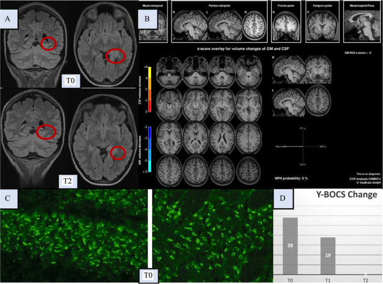
Fig. 1. Diagnostic findings initially (at T0) and after immunotherapy (at T1/T2).
Here, the magnetic resonance imaging (MRI) findings of the brain, the autoantibody findings, and the clinical course based on the Yale-Brown Obsessive-Compulsive Scale (Y-BOCS) scores are shown. Brain [18F]fluorodeoxyglucose positron emission tomography (FDG-PET) identified unremarkable findings, and no evidence of malignancy was identified in the whole body FDG-PET. Optical coherence tomography and electroencephalography were normal (these normal findings are not shown). A The left upper row shows the conspicuous FLAIR MRI findings of the brain with a solitary lesion in the left tapetum (at T0), atypical for age in location and localization and potentially (post)inflammatory, but the MRI criteria for multiple sclerosis were not met. In addition, a pineal gland cyst was found loco typico without a space-occupying effect (max. 11 mm in diameter; not shown in detail). The lower images show a regressive lesion in the left tapetum after clinical full remission (at T2, approximately 7 weeks after steroid pulse treatment). B A combined volume- and region-based analysis method (CVR; https://www.veobrain.com/?page=veomorph) detected no atrophy at T0. At time point T2, there was no relevant change (not shown). C In the cerebrospinal fluid (CSF), autoantibodies against granule cell cilia were found in the hippocampus (left in the bottom row; “rings and rods” pattern) and in neurons of the cortex (right in the bottom row; at T0). In addition, several large vessels were stained (not shown). The tissue-based assay using serum material revealed non-specific findings (not shown). The well-characterized anti-central nervous system autoantibodies against intracellular antigens (Yo/Hu/CV2/CRMP5/Ri/Ma1/2/SOX1/Tr/Zic4/GAD65/amphiphysin) or glial structures (AQP4/MOG) in serum and against cell surface antigens (NMDA-R/LGI1/CASPR2/AMPA1/2-R/GABA-B-R/DPPX) in serum and CSF remained unremarkable (not shown). Additional CSF analyses identified pleocytosis, with a white blood cell count of 10/µl (reference <5/µl); intrathecal immunoglobulin (Ig) synthesis of two isotypes, IgG (53%) and IgA (48%; reference both <10%); an elevated IgG index of 1.37 (reference <0.7); and CSF-specific oligoclonal bands. In contrast, the albumin quotient was normal, and pathogen testing of the CSF remained unremarkable. Elevated specific antibody indices (AIs) for HSV (2.3; reference <1.5) and VZV (2.8; reference <1.5) suggested polyclonal IgG synthesis, but the MRZ reaction remained negative (i.e., AIs for measles and rubella were negative). D The patient showed an initial Y-BOCS score of 29 points (at T0). At T1, immediately after steroid pulse treatment, the patient was able to better distance herself from the obsessive-compulsive symptoms (OCS), and the Y-BOCS score decreased to 19 points. Full remission of OCS (Y-BOCS score: 0) was evident at T2 (approximately 7 weeks after steroid pulse treatment). Neuropsychological testing of attentional performances revealed an improvement in reaction time (alertness with/without warning tone) at the time of the second assessment (directly after stopping steroids; not shown).

