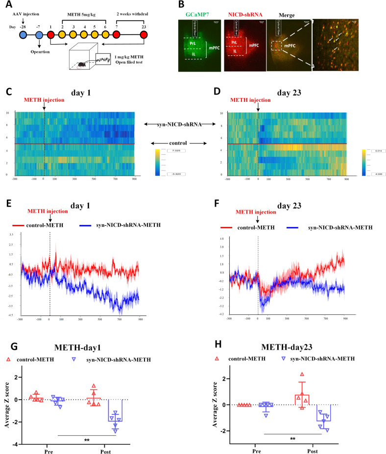
Fig. 3. Inhibition of the Notch1 signalling attenuates mPFC neuronal activity of METH-induced sensitized mice.
A Experimental design for recording GCaMP activity from mPFC neurons which reduced the Notch1 signalling during METH sensitization. Calcium-dependent (470 nm) and calcium-independent (410 nm) fluorescence signals were recorded before and after administration of METH (1 mg/kg) in the open field. B Fluorescence images in the mPFC from mice that received unilateral infusion of syn-GCaMP7f (green) and syn-NICD-shRNA (red) virus, scale bar = 100 µm. Arrows indicate neurons both expressed GCaMP7f and NICD-shRNA signals, scale bar = 40 µm. C, D Heatmap illustration of Ca2+ signals aligned to the initiation of trials on day 1 and day 23. Each row plots one trial, and a total of 10 trials were illustrated. The colour scale on the right indicates Z scores. E, F Average traces of calcium signals from control mPFC neurons (red line) and inhibitory NICD mPFC neurons (blue line) were recorded before (5 min) and after METH treatment (15 min) on days 1 and 23. G, H The average Z scores from (E) and (F) in the open field before (pre) and after (post) METH injection on day 1 and day 23. The fiber photometry signal of mPFC neurons in the syn-NICD-shRNA-METH group was significantly reduced compared with the baseline signal on day 1 and day 23, according to a paired-sample t test (t day1 = 7.953, t day23 = 4.58, **P < 0.01 vs. before METH injection). Traces represent mean ± SEM (E, F). Error bars represent mean ± SEM (G, H), n = 5.

