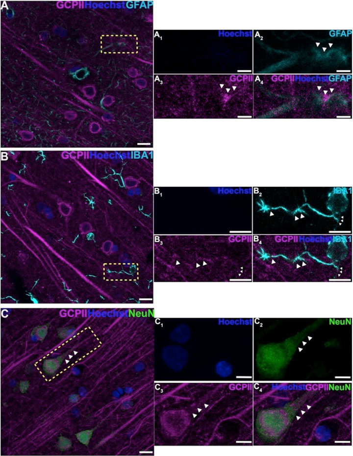
Fig. 2. GCPII cellular labeling in layer III of the aged monkey dlPFC using multiple label immunofluorescence.
A Another example of GCPII labeling (magenta) in astrocytes that are co-labeled with GFAP (cyan). (A1-A4) correspond to the yellow boxed region in (A). White arrowheads demarcate co-localization of GFAP and GCPII. B GCPII is occasionally observed in the processes of microglia, labeled with iba1 (cyan), and outlined by the white arrowheads. C Another example of GCPII co-localized in neurons (NeuN, green) with a pyramidal cell-like morphology. White arrowheads highlight GCPII labeling in an apical dendrite. Hoechst label (blue) indicates nuclei. (B1-B4), and (C1-C4) magnified regions correspond to the areas delineated by the yellow boxes in (B) and (C). Scale bars: (A–C) 10 μm; (A1-A4) 2.5 μm; (B1-B4) 5μm; (C1-C4) 5 μm.

