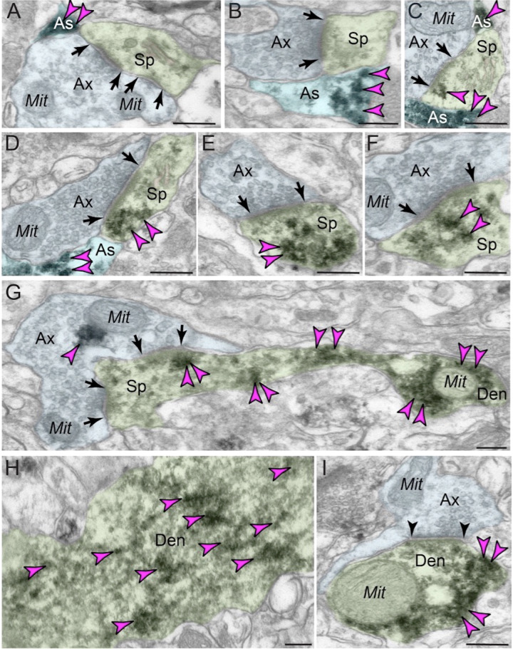
Fig. 3. ImmunoEM subcellular localization of GCPII in aged macaque dlPFC layer III.
A–C Immunoperoxidase labeling for GCPII in aged macaque (28–30 y) dlPFC layer III is visualized within astroglial leaflets ensheathing asymmetric (likely glutamatergic) synapses. GCPII immunolabeling appears to show a preference for the astroglial leaflets (PAPs) near the plasma membrane in perisynaptic locations near the excitatory glutamatergic synapse. D–G GCPII immunolabeling is prominently expressed in postsynaptic compartments in dendritic spines, near the plasma membrane and PSD synaptic active zone (D, F and G). Intriguingly, GCPII immunolabeling is visualized in vesicular-like structures within the dendritic spine head, positioned to undergo endocytosis or exocytosis (E and F). In (G), a dendritic spine is seen emanating from the dendritic shaft of a pyramidal cell; both are immunopositive for GCPII. Sparse presynaptic GCPII labeling is also observed in the axon terminal in association with mitochondria. All dendritic spines receive axospinous Type I asymmetric glutamatergic-like synapses, and the spine apparatus is pseudocolored in pink in the spine head. Synapses are between arrows. H–I In aged macaque dlPFC layer III, GCPII immunolabeling was visualized in dendritic shafts and was associated with microtubules oriented in parallel bundles. In (I), the dendritic shaft receives an axodendritic symmetric Type II synapse (arrowheads) and GCPII immunolabeling within the dendritic shaft was in close proximity to mitochondria. Color-coded arrowheads (magenta) point to GCPII immunoreactivity. Profiles are pseudocolored for clarity. Ax axon, Mit mitochondria, Sp dendritic spine, As astroglia, Den dendrite. Scale bars: 200 nm.

