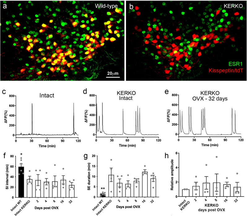
Fig. 4. GnRH pulse generator activity in Kiss1-specific ESR1 knockout (KERKO) mice.
a, b Representative examples of dual-label immunofluorescence for tdT (kisspeptin) and ESR1 (green) in wild-type (a, N = 4) and KERKO (b, N = 5)) mice. Almost all tdT neurons co-express ESR1 (yellow nuclei) while none occur in the KERKO mouse. c Representative example of GCaMP6 photometry recordings from an intact mouse wild-type at the Esr1 locus. d, e photometry recordings from KERKO mice when intact and 32 days after OVX. f–h Graphs showing mean ± SEM and individual data points for photometry parameters recorded from intact WT mice and KERKO mice before (intact) and 2, 4, 8, 16, and 32 days following OVX. Intact WT versus Intact KERKO *P = 0.038 and **P = 0.010 (Mann–Whitney test; N = 4)). No parameters changed at any time point after OVX (P > 0.05, one-way ANOVA, see text for exact values). Source data are provided as a Source Data file.

