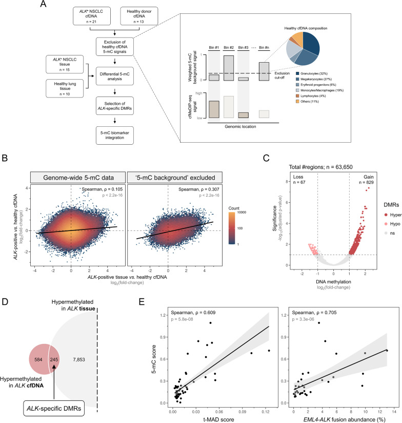
Fig. 1.
Identification of tumor-informative 5-mC regions from ALK-positive cfDNA. A 5-mC biomarker identification workflow overview. B Density plot illustrating log2 5-mC differences between ALK-positive patient and healthy control cfDNA (y-axis) against ALK patient tissue versus healthy control cfDNA (x-axis). Healthy control cfDNA samples used as reference for log2(fold-change) calculation were split, using one half as reference for ALK cfDNA (n = 6) and the other for ALK tissue samples (n = 6). The left plot shows the correlation at all evaluable genomic regions (n = 2,596,067). The right one shows the correlation after exclusion of ‘5-mC background ‘signals (n = 63,650). C Volcano plot of 63,650 genomic regions remaining after ‘5-mC background ‘exclusion. Significantly hyper- and hypomethylated sites, comparing 21 ALK-positive to 13 healthy control cfDNA samples, are highlighted in dark and light red, respectively. D Overlap of ALK cfDNA hyper-DMRs and hyper-DMRs detected in ALK tissue. E Scatter plots showing the correlation between 5-mC scores calculated from 245 ALK-specific hyper-DMRs and t-MAD scores (left) and EML4-ALK fusion abundances (right). Points represent individual samples and black line denotes fitted linear regression model with 95% confidence interval. ALK, anaplastic lymphoma kinase; cfDNA, cell-free DNA; DMR, differentially methylated region; EML4, echinoderm microtubule-associated protein-like 4; ns, not significant; NSCLC, non-small cell lung cancer; t-MAD, trimmed median absolute deviation from copy number neutrality

