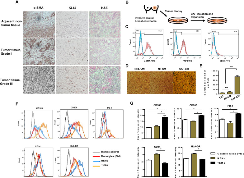
Fig. 1.
Stromal fibroblasts isolated from BC tissues exhibit characteristics of CAFs, effectively recruit monocytes and subsequently affect their polarization states. A Representative photomicrographs of H&E staining and IHC staining of α-SMA (a CAF-specific marker) and Ki67 (a proliferation marker) positive cells in breast tumor and adjacent non-tumor tissues (×100). Immunohistochemically, the presence of α-SMA demonstrated the presence of stromal myofibroblasts surrounding the cancer nests, which were distributed among the invasive cancer cells in the heterogeneous cancer tissue. B A schematic illustration of primary CAF isolation and expansion in vitro. C Flow cytometry staining of primary cultured CAFs with anti-α-SMA, anti-FAP, and anti-CD31. CAFs were characterized by the expression of α-SMA and FAP but lacked the expression of endothelial cell marker CD31 by flow cytometry. D, E To elucidate that monocytes are functionally recruited by CAFs, serum-starved monocytes were allowed to migrate for 12 h toward CMs from CAFs or NFs and compared to the negative control. D Representative photomicrographs of the migration potential of monocytes in different conditions assessed using the transwell assay. E Quantitative assessment of migrated cells showed that monocytes were effectively recruited by CAF-CM. Representative flow cytometry histograms (F) and bar graphs of mean fluorescent intensity (G) show that the expression levels of CD163, CD206, and PD-1 were considerably higher in TEMo than in NEMo or control monocytes, while the expression levels of HLA-DR and CD14 were significantly lower in TEMo compared to NEMo. Columns, mean of three different experiments; bars, SD. *P-value < 0.05, ***P-value < 0.001

