Fig. 4.
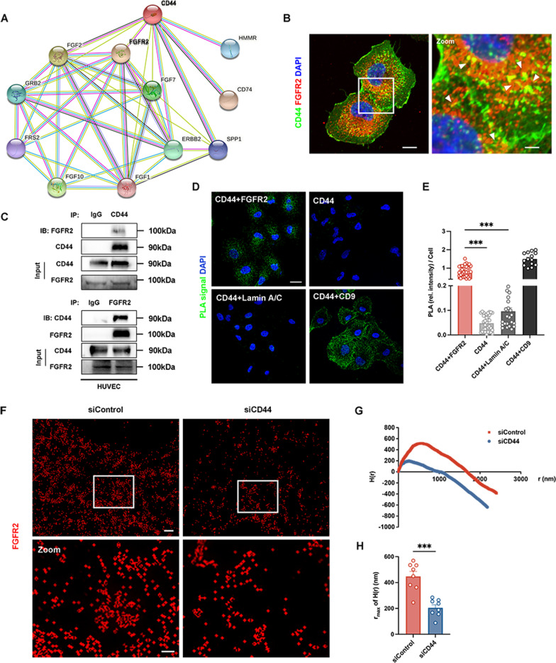
CD44 is associated with FGFR2 in ECs. A The STRING database was used to predict the protein-protein interaction network between CD44 and FGFR2. Pink edges represent the known protein-protein associations that are experimentally determined. Yellow edges are meant to be the predicated association via text mining. B Representative confocal microscopy images of CD44 (green) and FGFR2 (red) in HUVECs. Nuclei were stained with DAPI (blue). White arrowheads denote the colocalization of CD44 and FGFR2. Scale bars: 10 μm (left panel); 3 μm (right panel). C CD44 and FGFR2 were co-expressed in HUVECs, when the CD44 protein was immunoprecipitated by an anti-CD44 antibody, the indicated proteins, including FGFR2, were detected by immunoblotting. The same was true for the immunoprecipitation of the FGFR2 protein via an anti-FGFR2 antibody. Normal rabbit IgG acted as a negative control. D Representative confocal microscopy images of proximity ligation assay (PLA) signals (green) in HUVECs. CD44 served as the blank control (n = 32), CD44 + Lamin A/C served as the negative control (n = 22), and CD44 + CD9 served as the positive control (n = 14). CD44 + FGFR2 (n = 32). Scale bar: 20 μm. E The statistical data of D. F The FGFR2 distribution and clustering on the HUVEC cell surface were imaged at a nanoscale by N-STORM super-resolution microscopy. Scale bars: 1 μm (upper panel); 40 nm (lower panel). G Ripley’s H functions were acquired by NIS-Elements AR software. H The statistical data of the rmax of H(r) between the siControl and siCD44 groups. n = 8 per group. ***P < 0.001

