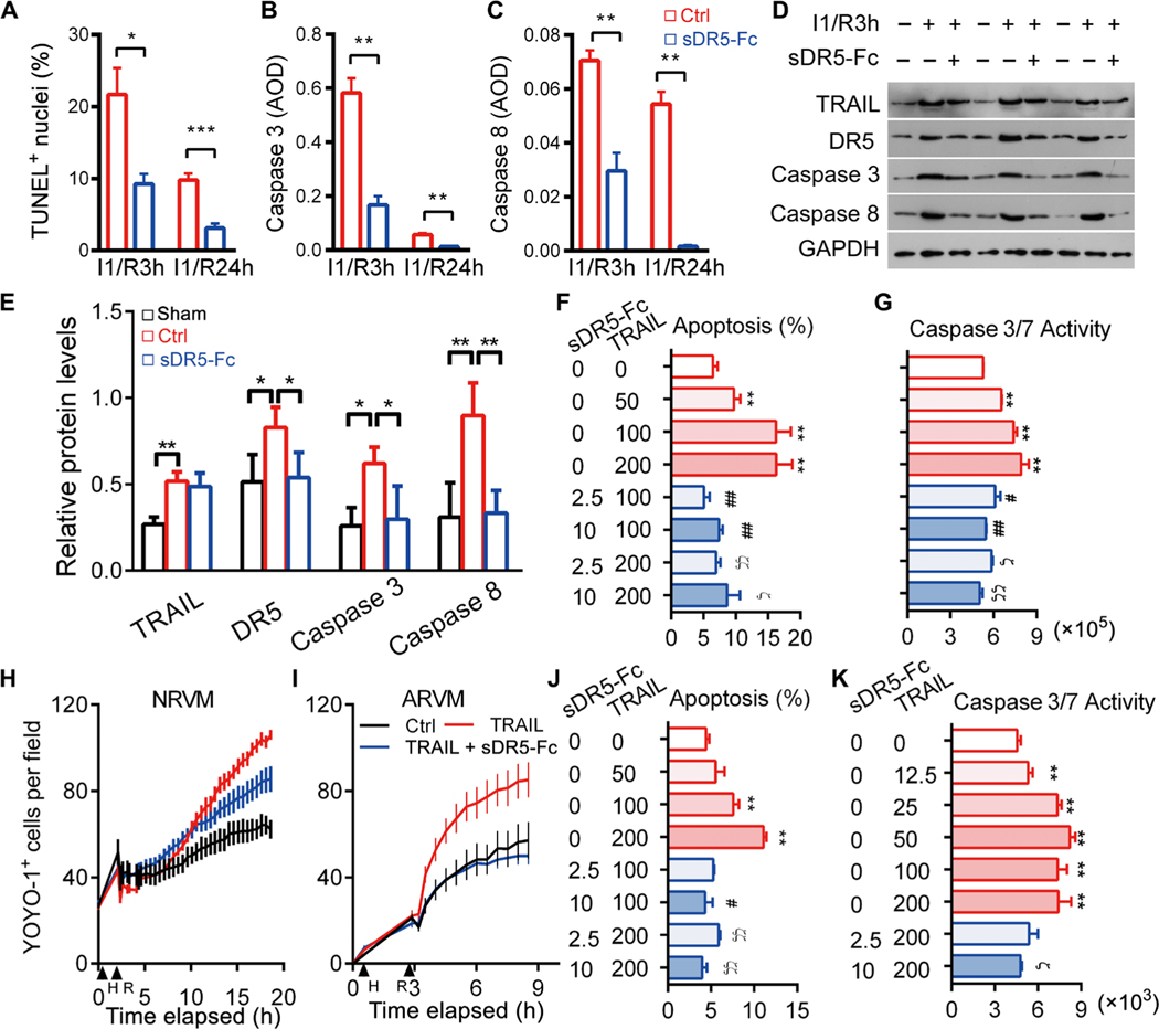
Fig. 5. sDR5-Fc protects rat and human cardiomyocytes from TRAIL-induced death.
(A) Percent TUNEL+ nuclei in rat heart sections after I1/R3h or I1/R24h with or without sDR5-Fc treatment (n ≥ 3 mice). (B and C) Caspase 3 (B) and caspase 8 (C) expressions as determined by IHC of heart sections described in (A) (n ≥ 3 per group). AOD, average optical density. (D and E) Immunoblot micrographs (D) and quantification (E) of rat heart homogenates after I1/R3h or I1/R24h with or without sDR5-Fc treatment, for DR5, TRAIL, activated caspase 3, activated caspase 8, and glyceraldehyde-3-phosphate dehydrogenase (GAPDH). Each lane represents one rat (n ≥ 3 rats). (F) Percent apoptotic cells as determined by TUNEL staining in NRVM cultures treated with TRAIL or sDR5-Fc as indicated under the H2/R3h stress condition. n ≥ 5 cultures. (G) Caspase 3/7 activity as determined by Caspase-Glo3/7 assay presented as relative luminescence unit (RLU) in cultures described in (F). (H and I) Number of dead cells per field of NRVMs and ARVMs treated with or without TRAIL or sDR5-Fc as determined by YOYO-1 dye staining and time-lapse microscopy with 30-min intervals. (J and K) Percent apoptotic cells as determined by TUNEL (J) and caspase 3/7 activity by Caspase-Glo3/7 assay (K) in human cardiac myocyte cultures treated with TRAIL or sDR5-Fc as indicated under the H3/R3h stress condition (n ≥ 3 cultures). Data are shown as means ± SEM. Statistical significance was determined by two-tailed t test (A to C) or one-way ANOVA followed by Tukey’s post hoc test (E to G and J to K). *P < 0.05, **P < 0.01, #P < 0.05, and ##P < 0.01 versus TRAIL group (100 ng/ml); ϚP < 0.05 and ϚϚP < 0.01 versus TRAIL group (200 ng/ml).

