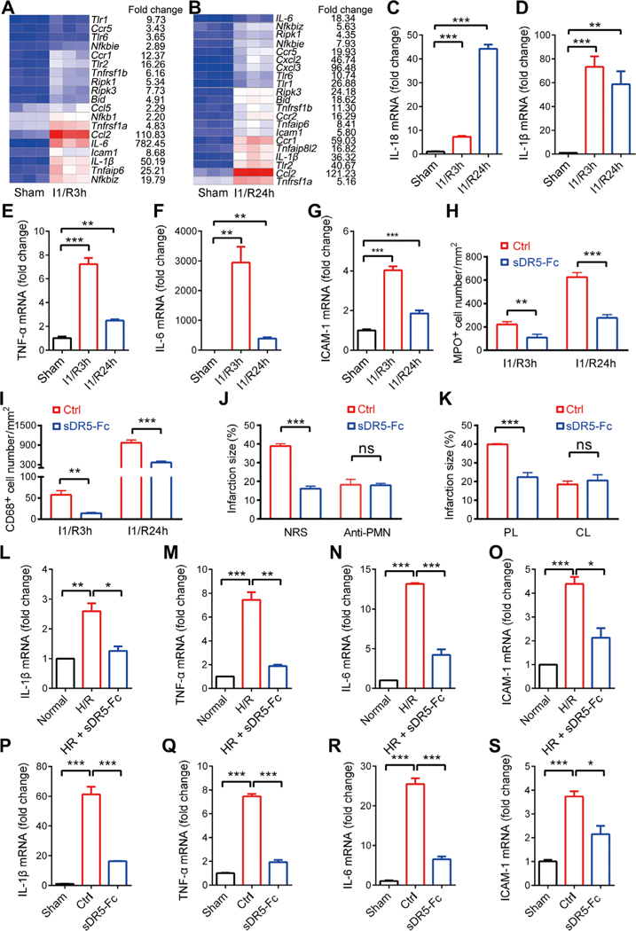
Fig. 6. The effect of sDR5-Fc on cardiac I/R is dependent on myeloid cells.
(A and B) Heat map of RNA-seq data of rat heart tissue from sham or ischemic animals. Each column represents three rats. (C to G) mRNA expression of IL-18, IL-1β, TNF-α, IL-6, and ICAM-1 in the myocardium of sham or ischemic rats as detected by quantitative PCR. (H and I) Numbers of MPO+ (H) and CD68+ (I) cells per mm2 of rat heart sections after the indicated treatments as determined by IHC (n ≥ 3 rats). (J) Infarct size as a percentage of the left ventricle in rats pretreated with a neutrophil-depleting antibody or normal rabbit serum (NRS) followed by I1/R24h with or without sDR5-Fc injection (5 min before reperfusion; n ≥ 4 rats). (K) Infarct size as a percentage of the left ventricle in rats pretreated with macrophage- depleting clodronate liposomes (CL) or control liposomes (PL) followed by I1/R24h with or without sDR5-Fc injection (5 min before reperfusion; n ≥ 4 rats). (L to O) mRNA expression of proinflammatory molecules in rat macrophage cell line NR8383 after hypoxia for 1 hour followed by reoxygenation for 3 hours, with or without sDR5-Fc during both hypoxia and reoxygenation, as determined by real-time PCR (n = 3) cultures. (P to S) mRNA expression of proinflammatory molecules in isolated cardiac infiltrating myeloid cells of rats after I1/R24h with or without sDR5-Fc administration (5 min before reperfusion), as determined by real-time PCR (n = 3 mice). Data are shown as means ± SEM. *P ≤ 0.05, **P ≤ 0.01, and ***P ≤ 0.001 as determined by two-tailed t test (I to K) or one-way ANOVA followed by Tukey’s post hoc test (C to H and L to S). The experiments were repeated at least three times with similar results.

