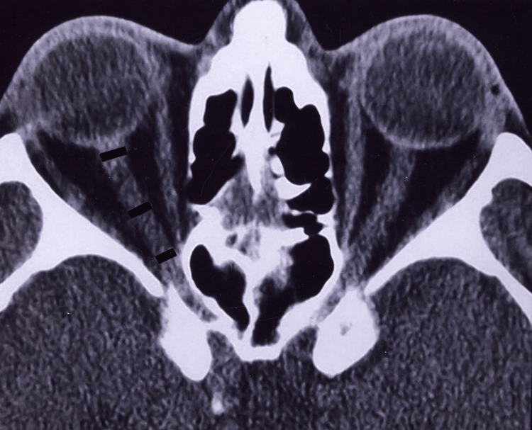
Figure 1. A patient with axial proptosis without glaucoma (normal retrobulbar optic nerve).
This figure shows the diameters of the retrobulbar optic nerve by axial computed tomography scan at the midglobe level in a 58-year-old man with bilateral axial proptosis without any clinical sign of glaucomatous optic nerve damage (visual acuities were 20/20 bilaterally, cup:disc ratio was 0.2 in both eyes, and visual field analysis demonstrated normal findings). Both measurements in the middle portion of the nerve (4.88 mm on the right and 5.00 mm on the left) were above the mean values of group 1 (mean, 3.49 mm) and group 2 eyes (mean, 3.77 mm) of glaucoma subjects.

