Figure 3.

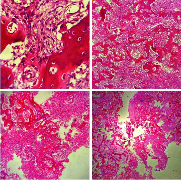
Histopathological images showed stroma containing osteoblasts, scattered dilated vascular channels, and large sheets of irregular trabeculae in osteoid material rimmed by osteoblast with ample cytoplasm and hyperchromatic nuclei

Histopathological images showed stroma containing osteoblasts, scattered dilated vascular channels, and large sheets of irregular trabeculae in osteoid material rimmed by osteoblast with ample cytoplasm and hyperchromatic nuclei