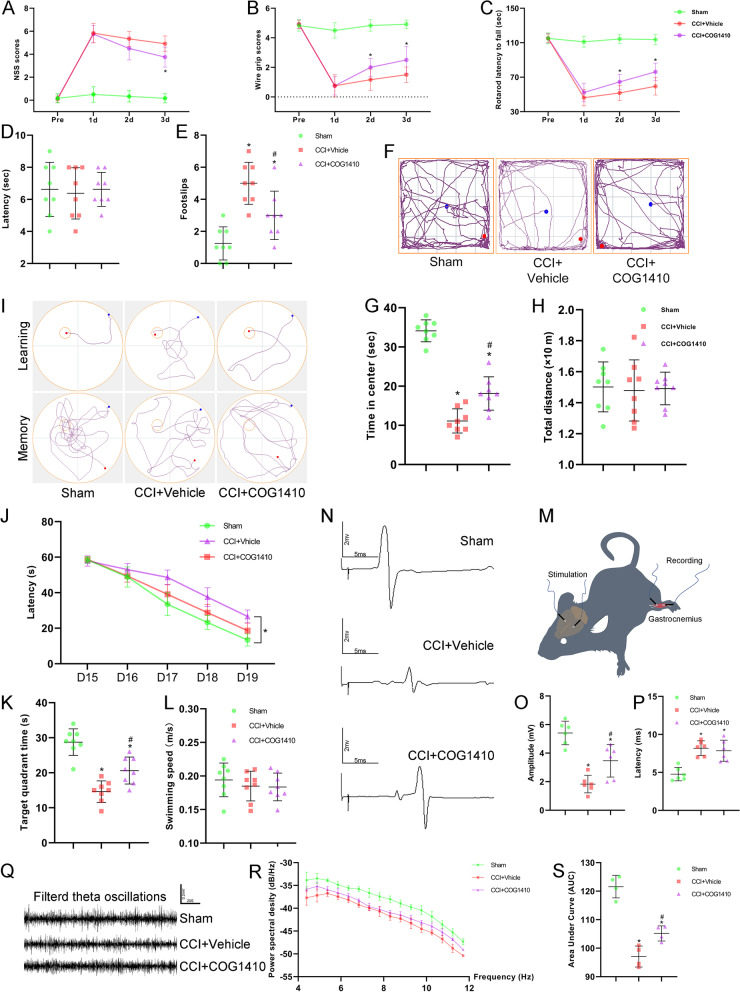
Fig. 3.
COG1410 treatment promoted neurological function and brain electrophysiological activity recovery. A–C Quantitative analysis of short-term neurological function by NSS scores (A), wire grip scores (B), and rotarod test (C). *p < 0.05 vs. CCI + Vehicle, n = 12 per group. D, E Quantitative analysis of motor function at 13 d after CCI by the beam walking test. *p < 0.05 vs. Sham; #p < 0.05 vs. CCI + Vehicle, n = 8 per group. F Representative images of the trajectory of mice in the open field test at 14 d after CCI. G, H Quantitative analysis of centre time (G) and total distance travelled by mice in the open field test (H). *p < 0.05 vs. Sham; #p < 0.05 vs. CCI + Vehicle, n = 8 per group. I Representative images of the trajectory of mice in the Morris water maze. J–L Quantitative analysis of latency in the learning test (J), target quadrant time (K), and average swimming speed in the target quadrant test (L). *p < 0.05 in J represents ANOWA; *p < 0.05 in K vs. Sham; #p < 0.05 in K vs. CCI + Vehicle, n = 8 per group. M Schematic diagram of motor evoked potential (MEP) monitoring in mice. N Representative images of MEP waves in different groups. O, P Quantitative analysis of MEP amplitude (O) and MEP latency (P). *p < 0.05 vs. Sham; #p < 0.05 vs. CCI + Vehicle, n = 6 per group. Q Representative images of filtered theta oscillations in different groups. R Representative power spectral density (PSD) curves of theta oscillations in different groups. S. Quantitative analysis of the total PSD of theta oscillations by the area under the curve (AUC). *p < 0.05 vs. Sham; #p < 0.05 vs. CCI + Vehicle, n = 4 per group

