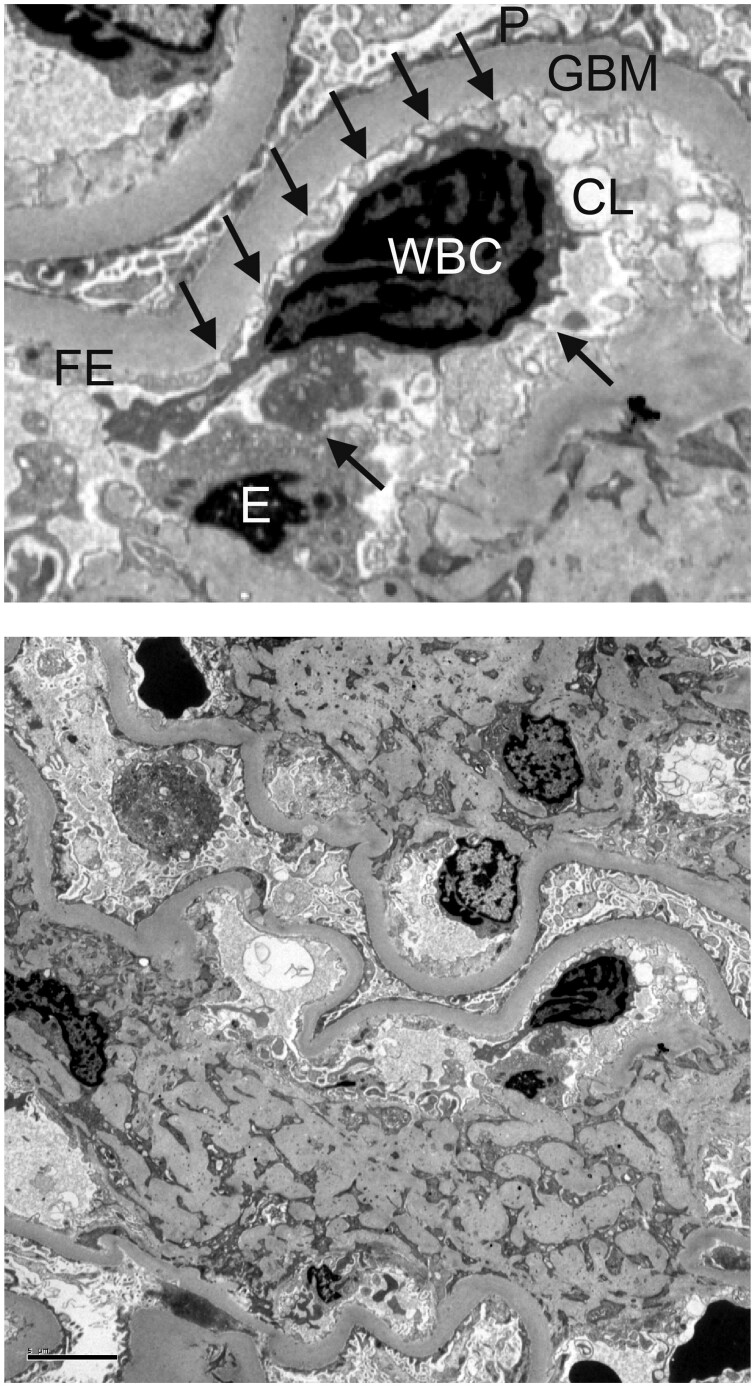
FIGURE 2.
Peripheral glomerular capillaries from a Pima Indian with Type 2 diabetes. In the upper panel, arrows point to projections extending from the WBC to the adjacent endothelium within the glomerular capillary lumen. Transmission electron microscopy ×3000. P, podocyte foot processes; E, endothelial cell body; FE, fenestrated endothelium; CL, capillary lumen. Complete image is shown in the lower panel. Scale bar illustrates a length of 5 µm.

