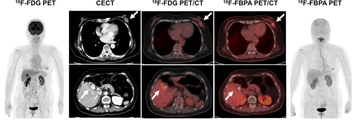
Fig. 5.
An 80-year-old woman underwent contrast-enhanced CT (CECT) for a recently diagnosed breast tumor before surgery. CECT revealed gallbladder wall thickening with gallstones and a contrast enhancement effect, in addition to a left mammary mass, which may indicate gallbladder cancer. 18F-FDG PET/CT showed a local increase in accumulation in the gallbladder wall (SUVmax: 8.1), making it difficult to differentiate gallbladder cancer from cholecystitis. 18F-FBPA PET/CT showed accumulation in the left breast mass (SUVmax: 4.5), but no significant accumulation in the gallbladder wall (SUVmax: 2.8). Cholecystectomy was performed and the pathological diagnosis was inflammatory granulation tissue

