
Figure 6. Novel electroporation-derived ccRCC syngeneic model is metastatic and transcriptomically resembles human stromal/proliferative ccRCC molecular subtype.
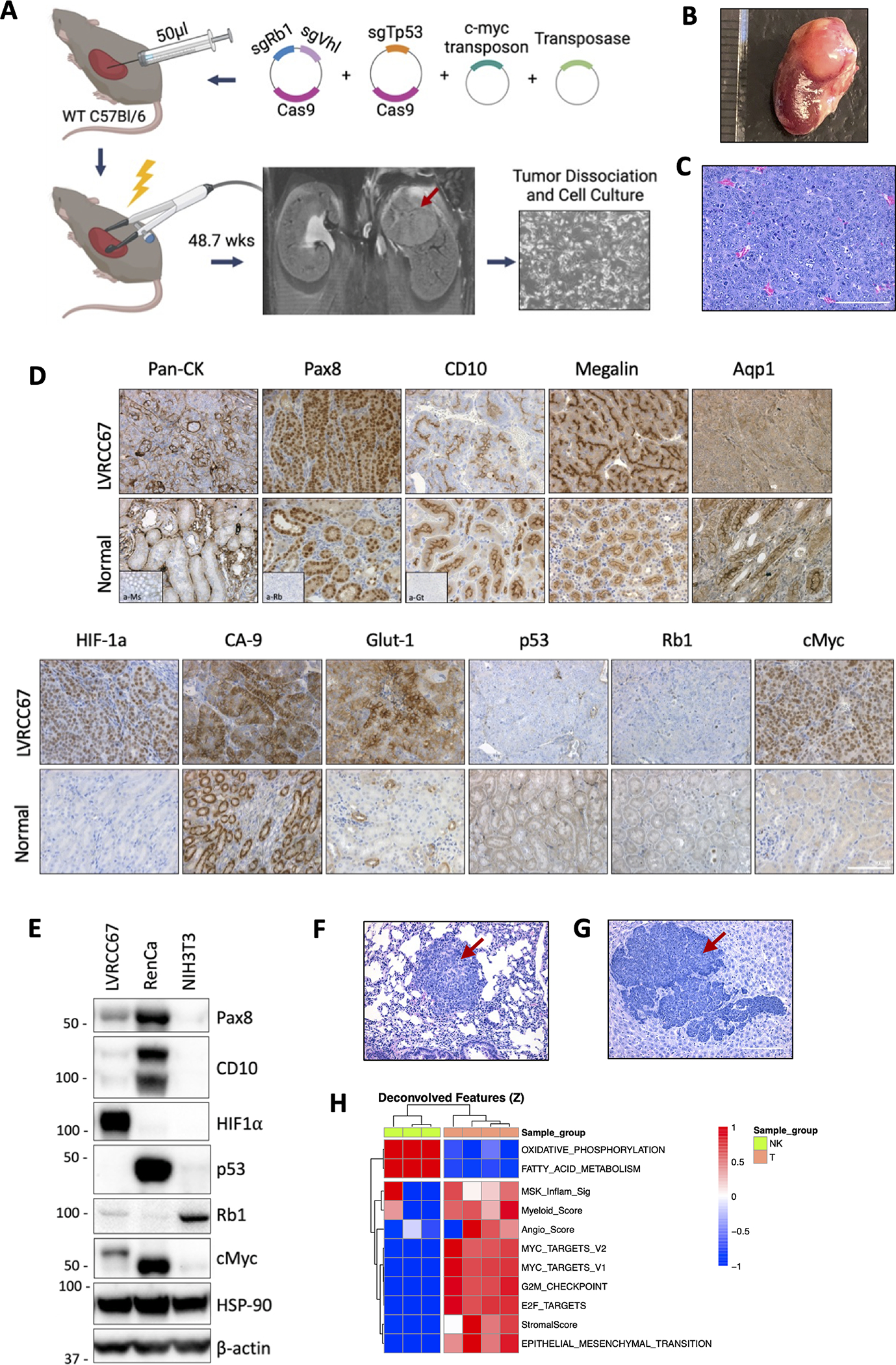
(A) Schematic of ccRCC syngeneic cell line development using electroporation of somatic tissues. Photo (B) and H&E (C) and IHC (D) of the parental kidney tumor and surrounding normal kidney. (E) Western blot of the EP-derived LVRCC67 cell line, RenCa and NIH3T3 cells are used as controls. H&E of lung (F) or liver (G) metastatic nodules following subcutaneous injection of LVRCC67 cells into WT C57Bl/6 mice. (H) Heatmap of ssGSEA scores in LVRCC67 tumors compared with normal kidney cortex samples from WT C57Bl/6 mice. Red arrows indicate tumor area. Scale bar 100μm. Schematic was made with BioRender.