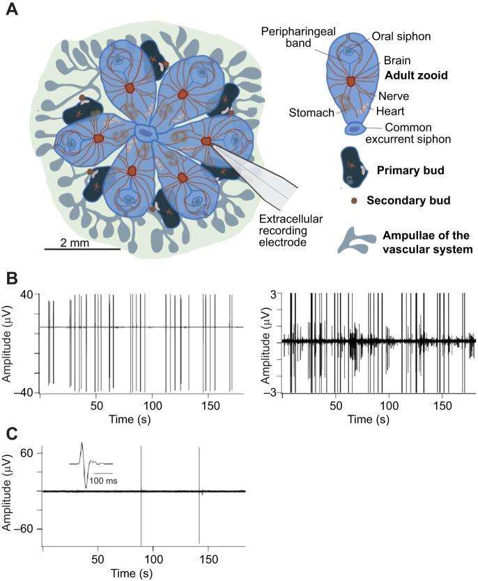
Fig. 1.
Brain activity at early and late developmental stages. (A) Illustration of a Botryllus schlosseri colony composed of a single module of six adult zooids with their associated primary and secondary buds, the location of the major organs, and the ampullae of the vascular network (credit: C. Anselmi). A recording electrode is shown positioned over the brain approximately to scale. (B) Extracellular recording from the brain of a mature zooid at developmental stage A3 showing spontaneous action potentials. The record is replotted at higher amplification to the right to emphasize the diversity of action potential waveforms. The amplitudes of the largest action potentials are truncated in the right panel. At this stage, the number of neurons is maximal (Anselmi et al., 2022). (C) Recording from the degenerating brain of a zooid approaching takeover (stage A5). Spontaneous activity is scarce and extracellular action potentials have a unitary waveform with little evidence of network activity. The inset shows an action potential on an expanded time base to illustrate the waveform. These results are representative of >100 recordings from >20 B. schlosseri clones.

