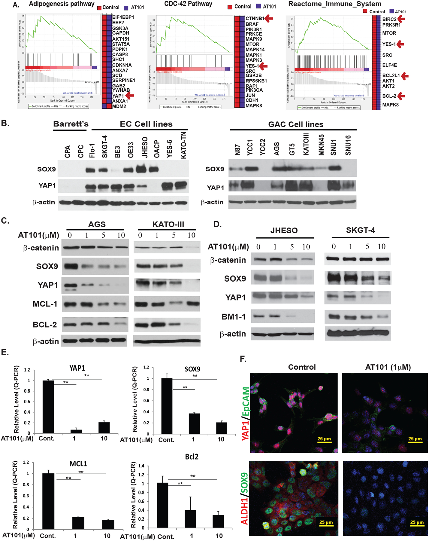
Figure 3. AT101 inhibits cell growth by suppressing MCL-1/BCL-2 and YAP/SOX9 axis.

A. RPPA proteomic analysis on AT101 treated AGS cells and the fold changes of genes affected by AT101 in AGC GC cells. B. Expression of YAP1 and SOX9 in two premalignant Barretts’ cell lines and eight EC cell lines and nine GC cell lines were detected by immunoblotting as described in Materials & Methods. Overexpression of both CSCs related genes YAP1 and SOX9 in EC and GC cell lines compared to premalignant cells. C. Down-regulation of antiapoptotic proteins-MCL-1/BCL2 and stem cell signaling-YAP/SOX9 and β-catenin in AGS and KATOIII cells treated with different dosage of AT101 determined by immunoblotting (left, middle panel). D. Down-regulation of proteins controlling stem cell signaling-YAP, SOX9, β-catenin and BMI-1 in both JHESO and SKGT-4 EAC cells treated with different dosage of AT101 by west blotting as indicated. E. AT101 strongly inhibits transcription of YAP1, SOX9, MCL-1 and BCL2 detected by real-time-PCR. **P<0.05. F. Expression of stem cell markers YAP1, EpCAM, SOX9 and ALDH1 were dramatically decreased upon treatment of AT101 at 1μM using confocal immunofluorescent staining in JHESO cells. Scale bar:25 μm.
