
Figure 4. AT101 sensitizes Docetaxel in inhibition of GC cell growth and promote apoptosis.
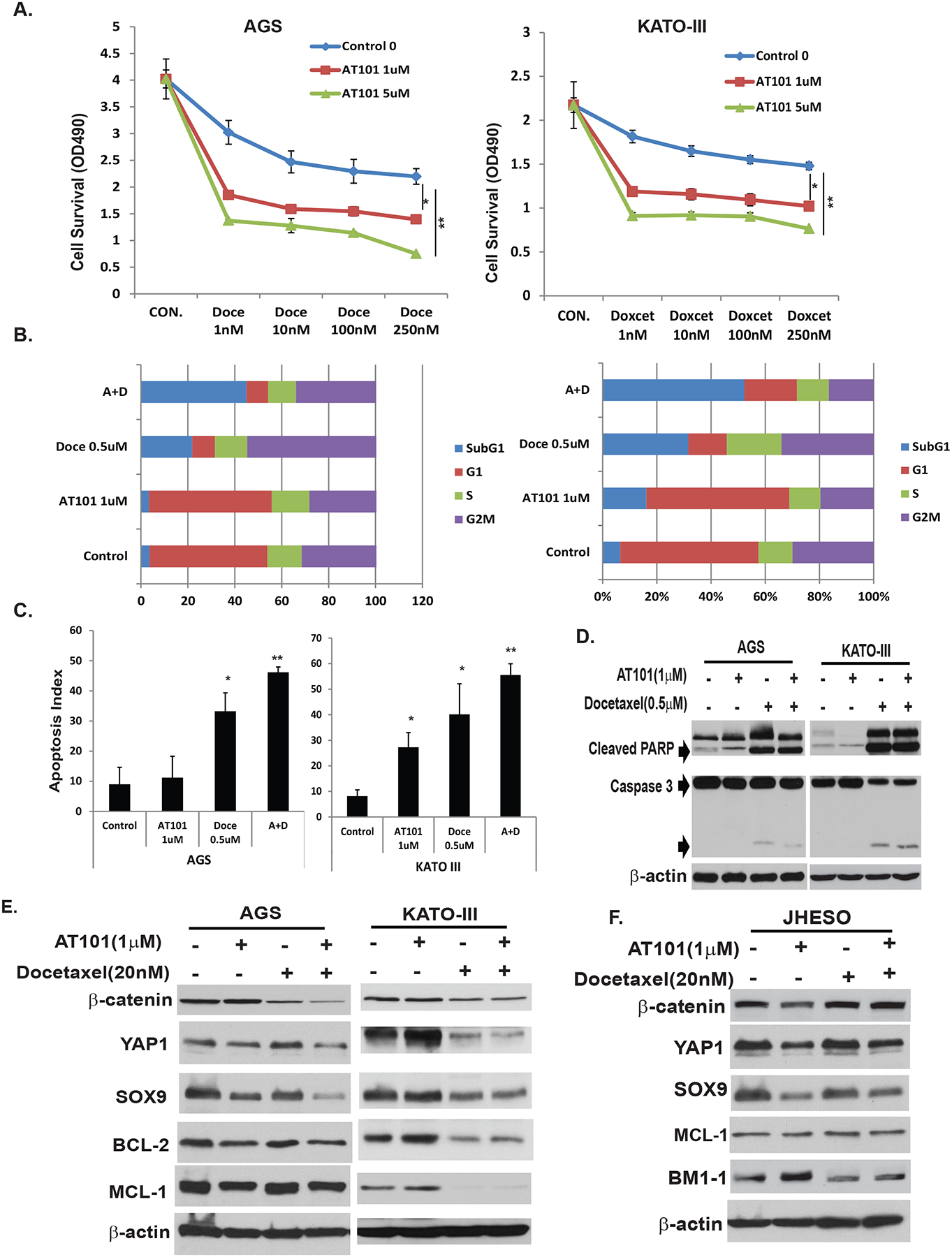
A. AGS and KATOIII GC cells were treated with docetaxel alone at dosage of 1nM to 250nM or in combination with AT101 at 1μM and 5μM respectively for 5 days, cell growth was determined using MTS as indicated in Materials and Methods. B. AGS and KATO-III cells were seeded onto 6-well plates and treated with 0.1% DMSO (as control) or with AT101 1μM or Docetaxcel 0.5μM or in combination for 48 hours and then fixed and stained for DNA with propidium iodide and then analyzed for DNA histograms and cell cycle phase distribution by flow-cytometry using a FACS Calibur. Results demonstrated that AT101 significantly sensitize docetaxel in inhibition of cell growth in a dose dependent manner and the combination of AT101 and docetaxel strongly increase cells in sub-G1. C. AGS and KATOIII cells were treated with 0.1% DMSO (as control) or AT101 or/and docetaxel at the dosage indicated, and then determined the apoptosis index by flow cytometry. Results indicated that the apoptosis index was significantly increased when cells treated with the AT101 in combination with docetaxel. D. Cell total lysates were isolated from AGS and KATO-III cells treated with 0.1% DMSO (as control) or AT101 or/and docetaxel at the dosage indicated for 48 hours, total and cleaved PARP and caspase 3 were determined by immunoblotting as described in material & Methods; E&F. Cell total lysates were isolated from AGS, KATO-III GC cells and JHESO EAC cells treated with 0.1% DMSO (as control) or AT101 or/and docetaxel at the dosage indicated for 48 hours, expression of stem cell signaling proteins-YAP1,SOX9 and β-catenin and antiapoptotic proteins MCL-1/BCL-2 were determined by immunoblotting as described in Materials & Methods.